Tag Archives: fracture

What Is: The Galeazzi Fracture?

Orthopedic surgeons have so many names for fractures, it gets confusing! Today, let’s dig in to the “Galeazzi fracture.” This one was named for an Italian surgeon during the early 20th century) although it was actually first described by an Englishman named Cooper a hundred years earlier).

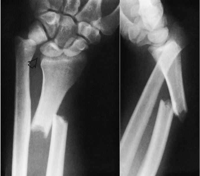
The Galeazzi fracture is an uncommon one, and consists of two components: a radius fracture at the junction of the distal and middle thirds, and a dislocation of the distal radio-ulnar joint. Here’s what it looks like:

Notice the obvious dislocation seen in the lateral view. Of course, a whole classification system has been developed to describe the various nuances of this fracture pattern, but that’s beyond the scope of this post.

What to do about it? This one needs prompt orthopedic consultation, and due to the dislocation component it requires operative management in adults. In children, initial closed reduction is the treatment of choice.

Monday, I’ll describe this fracture’s evil twin, The Monteggia fracture.

Trauma Mythbusters: NSAIDs And Fracture Healing

Trauma hurts like hell. Over the years, we’ve developed quite a few ways of combating this pain. A number of drug classes have been developed to reduce it. One of the more common non-narcotic drug classes are the NSAIDs. As I’ve mentioned before, every drug has dozens of effects. Drug companies market a particular medication based on one of the predominant effects. All the others are considered side effects.

NSAIDs are not unique; they have lots of side effects as well. In 2003, several papers brought to light possible interactions between these drugs and fracture healing. Specifically, there were questions about these drugs interfering with the healing process and of increasing the number of delayed unions or nonunions. But once again, how convincing were these papers, really?

It would seem to make sense that NSAIDs could interfere with bone healing. This  process relies heavily on the regulation of osteoblast and osteoclast function, which itself is regulated by prostaglandins. Since prostaglandins are synthesized by the COX enzymes, COX inhibitors like the NSAIDs should have the potential to impair this process. Indeed, animal studies in rats and rabbits seem to bear this out.

But as we have seen before, good animal studies don’t always translate well to human experience. Although a study from 2005 suggested that NSAID administration in older patients within 90 days of injury had a higher incidence of fracture nonunion, the study design was not a very good one. It is equally likely that patients who required these drugs in this age group may have been at higher risk for nonunion in the first place.

In fact, there are no large, prospective randomized studies that have explored the effect of short-term or long-term NSAID administration on fracture repair. But there have been several smaller studies that showed absolutely no effect on nonunion with short-term administration of this drug class. Yet the dogma that leads us to avoid giving these drugs persists.

A recent analysis looked at the quality of the published research, both for and against NSAID usage in fracture patients. They used the Coleman Methodology Score, which evaluates study size and type, mean followup, detailed description of treatment, subject selection, outcomes, and outcome assessment. The maximum score was 100. 

Here are the factoids:

  • There were 4x as many total subjects in the “NSAIDS are okay” papers than in the “avoid NSAIDS” papers
  • The quality of the “NSAIDS are okay” papers were significantly higher than “avoid NSAIDS” group (59 vs 40)
  • Interestingly, the “avoid NSAIDS” papers are cited twice as often
  • All of the reviews ended with my pet peeve catch phrase “further (good) research is needed”

Bottom line: Once again, the animal data is clear but the human data is not. Although there are theoretical concerns about their use, there is not enough solid risk:benefit information to abandon short-term NSAID use in patients who really need them. NSAIDs can and should be prescribed in patients with short-term needs and simple fractures.

References:

  1. Effects of nonsteroidal anti-inflammatory drugs on bone formation and soft-tissue healing. J AM Acad Orthop Surg 12:139-43, 2004.
  2. Effect of COX-2 on fracture-healing in the rat femur. J Bone Joint Surg Am 86:116-123, 2004.
  3. Effects of perioperative anti-inflammatory and immunomodulating therapy on surgical wound healing. Pharmacotherapy 25:1566-1591, 2005.
  4. Pharmacological agents and impairment of fracture healing: what is the evidence? Injury 39:384-394, 2008.
  5. High dose nonsteroidal anti-inflammatory drugs compromise spinal fusion. Can J Anaesth 52:506-512, 2005.
  6. Nonsteroidal Anti-Inflammatory Drugs and Bone-Healing: A Systematic Review of Research Quality. JBJS Rev 4(3), 2016.
Part 1: Metal Splints – Can You X-ray Through Them?

Splinting is an important part of the trauma resuscitation process. No patient should leave your trauma resuscitation room without splinting of all major fractures. It reduces pain, bleeding, and soft tissue injury, and can keep a closed fracture from becoming an open one.

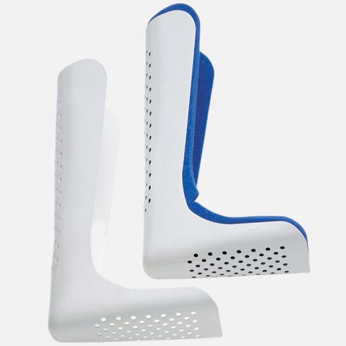
But what about imaging? Can’t the splint degrade x-rays and hamper interpretation of the fracture images? Especially those pre-formed aluminum ones with the holes in them? It’s metal, after all.

Some of my orthopedic colleagues insist that the splint be removed in the x-ray department before obtaining images. And who ends up doing it? The poor radiographic tech, who has no training in fracture immobilization and can’t provide additional pain control on their own.

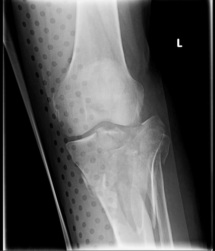
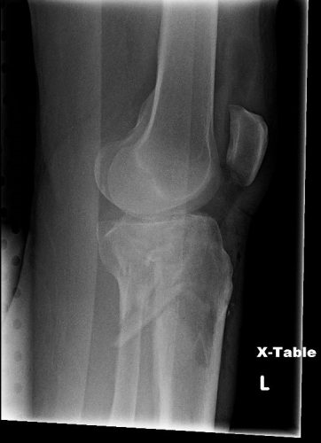
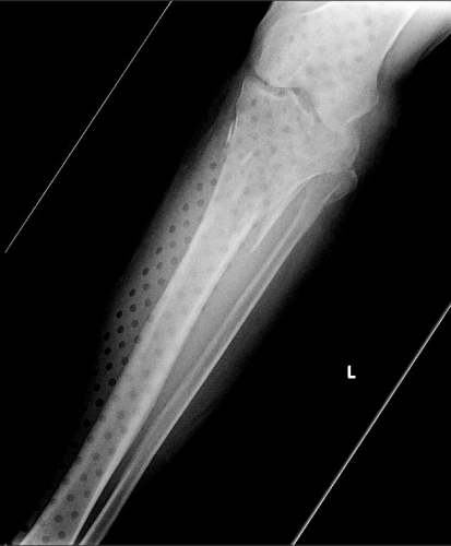
But does it really make a difference? Judge for yourself. Here are some knee images with one of these splints on:

Amazingly, this thin aluminum shows up only faintly. There is minimal impact on interpretation of the tibial plateau. And on the lateral view, the splint is well posterior to bones.

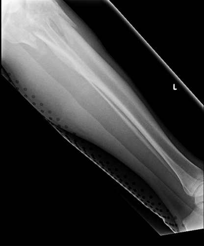
On the tib-fib above, the holes are a little distracting on the AP view, but still allow for good images to be obtained.

Bottom line: In general, splints should not be removed during the imaging process for acute trauma. For most fractures, the images obtained are more than adequate to define the injury and formulate a treatment plan. If the fracture pattern is complex, it may be helpful to temporarily remove it, but this should only be done by a physician who can ensure the fracture site is handled properly. In some cases, CT scan may be more helpful and does not require splint removal. And in all cases, the splint should also be replaced immediately at the end of the study.

In my next post, I’ll look at the use of CT scans when this type of splint is in use.


What You Need To Know About Frontal Sinus Fractures

Fracture of the frontal sinus is less common than other facial injuries, but can be more complex to deal with, both in the shorter and longer terms. These are generally high energy injuries, and facial impact in car crashes is the most common mechanism. Fists generally can’t cause the injury, but blunt objects like baseball bats can.

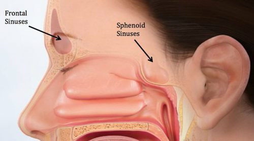
Here’s the normal anatomy:

sinus-fracture-treatment


Source: www.facialtraumamd.com

There are two “tables”, the anterior and the posterior. The anterior is covered with skin and a small amount of subcutaneous tissue. The posterior table is separated from the brain by the meninges.

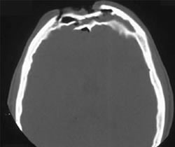
Here’s an image of an open fracture involving both tables. Note the underlying pneumocephalus.

frontal_sinus1

A third of injuries violate the anterior table, and two thirds violate both. Posterior table fractures are very rare. A third of all patients will develop a CSF leak, typically from their nose.

These fractures may be (rarely) identified on physical exam if deformity and flattening is noted over the forehead. Most of the time, these patients undergo imaging for brain injury and the fracture is found incidentally. Once identified, go back and specifically look for a CSF leak. Clear fluid in the nose is, by definition, CSF. Don’t waste time on a beta-2 transferring (see below).

If a laceration is clearly visible over the fracture, or if a CSF leak was identified, notify your maxillofacial specialist immediately. If more than a little pneumocephalus is present, let your neurosurgeon know. Otherwise, your consults can wait until the next morning.

In general, these patients frequently require surgery for the fracture, either to restore cosmetic contours or to avoid mucocele formation. However, these are seldom needed urgently unless the fracture is an open fracture with contamination or there is a significant CSF leak. If in doubt, though, consult your specialist.

Related posts:

EAST 2018 #7: Cervical Spine Injury And Dysphagia

One of the under-appreciated complications of cervical spine fractures is dysphagia. This problem disproportionately affects the elderly, and is most common in patients with C1-C3 fractures. Swallowing becomes even more difficult when the head is held in position by a rigid cervical collar, which is the most common treatment for this injury.

How common is dysphagia in patients with cervical spine injury? What is the best way to detect it? These questions were asked by the group at MetroHealth Medical Center in Cleveland. They  retrospectively reviewed their experience with patients presenting with cervical spine injury for 14 months, then prospectively studied the use of routine, nurse-driven bedside dysphagia screening in similar patients for a year. They wanted to test the utility of screening, and judge its impact on outcome.

Here are the factoids:

  • 221 patients were prospectively studied and received a bedside dysphagia screen, but only 114 met all inclusion criteria and had the protocol properly followed (!)
  • 17% had dysphagia overall, with an incidence of 15% in cervical spine injuries and 31% in those with a concomitant spinal cord injury
  • The bedside dysphagia screen was 84% sensitive, 96% specific, with positive and negative predictive values of 80% and 97%, respectively
  • There were 6/214 patients with dysphagia complications in the retrospective group vs 0/114 in the screened group

Bottom line: This abstract actually puts a number on the incidence of dysphagia on this group of patients. I wish the patient numbers could have been higher, but they are still very good. The results are convincing, and the negative predictive value is excellent. If the screen is passed, then the patient should do well with feeds. I recommend that all patients with cervical spine injury treated with a rigid collar undergo this simple screen, and have appropriate diet adjustments to limit complications.

Here are some questions for the authors to consider before their presentation:

  • Please share the details of the nurse-driven component of the bedside dysphagia screen, and how you determine when a formal barium swallow is indicated
  • Why did your prospective study group drop from 221 to 114?
  • When did you typically perform the screen? Fracture swelling may not peak for 3 days, so early screening may not be as good as later screening.
  • This was a nice study, with a very practical and actionable result!

Reference: EAST Podium abstract #10.