Category Archives: Head

The Modified Brain Injury Guideline Criteria (mBIG)

In my previous post, I reviewed the Brain Injury Guideline criteria (BIG) that were published in 2014, and cited some early papers promoting its use for simplifying neurotrauma care. These criteria allowed trauma professionals to use our neurosurgical colleagues’ services more sensibly.

As a reminder, these are the original BIG criteria:

Some revisions were proposed in 2020 to improve patient safety and reproducibility further.  Here are the revised criteria:

So, what are the differences?

  • The “loss of consciousness” was changed to a more objective assessment, the GCS
  • Intoxication is defined as a blood alcohol concentration > 80 mg/dL
  • Aspirin and NSAIDs are not considered antiplatelet agents
  • Epidural hematoma (EDH) is no longer sized; any epidural moves the patient to mBIG 3
  • Multiple intraparenchymal hemorrhages (IPH) move the patient to mBIG 3
  • Subarachnoid hemorrhage is more objectively classified

The mBIG criteria were tested in a multi-institutional review comparing the original criteria with the modified criteria. BIG 3 patients were excluded, since these patients required admission and neurosurgical consultation, which is maximal therapy. All patients underwent repeat CT scans to monitor for progression of the injury.

Here are the factoids:

  • A total of 269 patients were included; 98 were BIG 1 and 171 were BIG 2
  • In both BIG 1 and BIG 2 cohorts, CT progression was seen in about 11% of patients. These patients tended to have more severe injuries overall and were more likely to have EDH or IPH.
  • Two BIG 2 patients decompensated and required neurosurgical intervention; both had EDH

These findings prompted the changes that are now part of the mBIG score. Here is the complete algorithm based on the mBIG criteria (click to see full-size):

A larger validation study was published in 2022 by the same authors, following the addition of 496 patients from the same three trauma centers. The total number of patients included in the study was 496.

More factoids:

  • There were now a total of 223 mBIG 1 patients and 273 mBIG2
  • The number of CT scans and neurosurgery consults was significantly decreased
  • Hospital length of stay was also significantly decreased

Bottom line: The mBIG criteria perform better and are at least as safe as the initial BIG criteria. The mBIG criteria are more objective, making it easier to stratify patients accurately. 

The mBIG criteria should be adopted by any center seeking a consistent and validated process for stratifying patients for observation, admission, or neurosurgical consultation following head trauma. This will conserve resources and allow our neurosurgical colleagues to focus on the patients who truly need them.

References:

  1. Multicenter assessment of the Brain Injury Guidelines and a proposal of guideline modifications. Trauma Surgery & Acute Care Open, 5(1), e000483.
  2. A multicenter validation of the modified brain injury guidelines: Are they safe and effective?. Journal of Trauma and Acute Care Surgery 93(1):p 106-112, July 2022.

The BIG Brain Injury Guidelines

Until five years ago, there was tremendous variability in the way brain injuries were managed at trauma centers. There were no clear guidelines describing what should be done during the initial evaluation, and no consensus as to when to involve neurosurgery. This resulted in a lot of flailing about and unnecessary calls to our neurosurgical colleagues.

Then the Brain Injury Guidelines (BIG) came along 15 years ago. They were developed to allow trauma programs to stratify head injuries in such a way as to better utilize resources such as hospital beds, CT scanning, and neurosurgical consultation.

Injuries are stratified into three BIG categories, and management is based on them. Here is the stratification algorithm:

And here is the management algorithm based on the stratification above:

(RHCT = repeat head CT)

The original study was published ten years ago and was a retrospective review of 4,000 patient records. It found that a significant number of these patients could be managed exclusively by the trauma surgeons.

The AAST BIG Multi-Institutional Group set about prospectively validating this system to ensure that it was accurate and safe. They identified adult patients from ten high level trauma centers that had a positive initial head CT scan. They looked at the the need for neurosurgical intervention, change in neuro exam, progression on repeat head CT, any visits to the ED after discharge, and readmission for the injury within 30 days.

Here are the factoids:

  • About 2,000 patients were included in the study, with BIG1 = 15%, BIG2 = 15%, and BIG3 = 70% of patients
  • BIG1: no patients worsened, 1% had progression on CT, none required neurosurgical intervention, no readmits or ED visits
  • BIG2: 1% worsened clinically, 7% had progression on CT, none required neurosurgical intervention, no readmits or ED visits
  • All patients who required neurosurgical intervention were BIG3 (20% of patients)

The authors concluded that using the BIG criteria, CT scan use and neurosurgical consultation would have been decreased by 29%.

Bottom line: This is an exciting paper! BIG has been around for awhile, and some centers have already started using it for planning the management of their TBI patients. This study provides some validation that the system works and keeps patients safe while being respectful of resource utilization. 

My only criticism is that the number of patients in the BIG1 and BIG2 categories is low (about 600 combined). Thus, our experience in these groups remains somewhat limited. However, the study is very promising, and more centers should consider adopting BIG to help them refine their management of TBI patients. 

This was the first prospective study of the BIG criteria. A great deal of additional work has been done. And now, an attempt has been made to simplify this algorithm even further.

In my next post, I’ll review the modified BIG (mBIG) criteria and describe them in detail.

References:

  1. The BIG (brain injury guidelines) project: defining the management of traumatic brain injury by acute care surgeons. Journal of Trauma and Acute Care Surgery, 76(4), 965-969.
  2. Validating the Brain Injury Guidelines: Results of an American Association for the Surgery of Trauma prospective multi-institutional trial. J Trauma Acute Care Surg. 2022 Aug 1;93(2):157-165.

Are Prophylactic Antibiotics Needed For Facial Fractures?

The use of prophylactic antibiotics in patients with facial fractures has been controversial since forever. Some trauma professionals argue that these fractures, many of which involve a sinus or the mouth, should be considered as open fractures.

Several studies on the use of antibiotics prophylactically, preoperatively, and postoperatively have shown a significant amount of variability. A few have shown no benefit from the use of short-, long-, or no antibiotics. In fact, the Surgical Infection Society issued a practice guideline on antibiotic use in facial fractures. Essentially, they recommended that antibiotics not be administered to patients who do not require surgery. And for operative fractures, they recommended against pre- or post-operative antibiotics.

A recently published study examined current practices regarding antibiotic administration, timing, and adverse events. The null hypothesis was that prophylactic antibiotics would not reduce facial fracture-associated infectious complications in nonoperative facial fractures.

The AAST Facial Fracture Study Group performed a prospective, observational study of adult patients who did not undergo operative repair of their facial fractures. Patients receiving antibiotics for other causes,  those who were immunocompromised, and patients with bowel injuries were excluded. The primary outcome was any related infection, drainage, or follow-up visit requiring antibiotics. Secondary outcomes included demographic indicators such as length of stay, ventilator time, discharge disposition, and readmission within 30 days.

Here are the factoids:

  • A total of 1,835 patients were studied, and two-thirds (64%) did not receive any antibiotics
  • Infections developed in 0.7% of patients without antibiotics and 1.7% with
  • The vast majority of fractures in all patients (84%) were not considered open (no mucosal exposure)
  • Antibiotic administration had a significant association with infectious complications, although the duration of antibiotics did not seem to make a difference

The authors concluded that infection rates were very low despite the majority of patients receiving no antibiotics.

Bottom line: This study provides another set of data points that show us that antibiotics are not necessary in many facial fractures. This is an observational study, so there were wide variations in practice patterns that make the study more difficult to interpret.

There was a relatively small number of patients with “open fractures” that involved exposure to the mucosa. This weakens the study conclusions for this group.

This study joins a growing number that would indicate that nonoperatively managed facial fractures do not require antibiotics. For those that do need surgery, the usual perioperative antibiotic rules still apply.

Reference: Prophylactic antibiotic use in trauma patients with non-operative facial fractures: A prospective AAST multicenter trial. Journal of Trauma and Acute Care Surgery 98(4):p 557-564, April 2025.

Everything You Wanted To Know About: Cranial Bone Flaps

Patients with severe TBI frequently undergo surgical procedures to remove clot or decompress the brain. Most of the time, they undergo a craniotomy, in which a bone flap is raised temporarily and then replaced at the end of the procedure.

But in decompressive surgery, the bone flap cannot be replaced because doing so may increase intracranial pressure. What to do with it?

There are four options:

  1. The piece of bone can buried in the subcutaneous tissue of the abdominal wall. The advantage is that it can’t get lost. Cosmetically, it looks odd, but so does having a bone flap missing from the side of your head. And this technique can’t be used as easily if the patient has had prior abdominal surgery.

2. Some centers have buried the flap in the subgaleal tissues of the scalp on the opposite side of the skull. The few papers on this technique demonstrated a low infection rate. The advantage is that only one surgical field is necessary at the time the flap is replaced. However, the cosmetic disadvantage before the flap is replaced is much more pronounced.

3. Most commonly, the flap is frozen and “banked” for later replacement. There are reports of some mineral loss from the flap after replacement, and occasional infection. And occasionally the entire piece is misplaced. Another disadvantage is that if the patient moves away or presents to another hospital for flap replacement, the logistics of transferring a frozen piece of bone are very challenging.

4. Some centers just throw the bone flap away. This necessitates replacing it with some other material like metal or plastic. This tends to be more complicated and expensive, since the replacement needs to be sculpted to fit the existing gap.

So which flap management technique is best? Unfortunately, we don’t know yet, and probably never will. Your neurosurgeons will have their favorite technique, and that will ultimately be the option of choice.

Reference: Bone flap management in neurosurgery. Rev Neuroscience 17(2):133-137, 2009.

What Is: A Hinge Fracture Of The Skull?

Although very few things in medicine are new, I love it when I learn about something I’ve never heard of before. Recently, while reading an autopsy report, I ran across the term “hinge fracture of the skull.” What? Maybe if I were a neurosurgeon, I would have recognized the term. This was the perfect excuse to hit the books (or, more accurately, the internet).

A hinge fracture crosses the skull base transversely and involves the temporal and sphenoid bones. Here are diagrams of two common transsphenoidal fracture patterns, courtesy of radiopaedia.org. The red and green lines can be considered transverse (hinge) fractures.

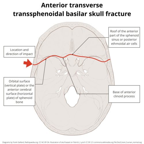
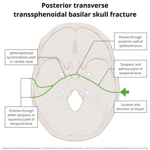
Why the hinge analogy? Since the fracture extends entirely across the skull base, it splits the skull in two. In theory, the bones could hinge around this line, but the reality is that it usually doesn’t. It’s just a memorable name.

It takes a significant amount of force to fracture the skull like this. Although any major blunt force could do this, there is a higher association with motorcycle crashes. I found an interesting paper (cited below) that showed that if a rider’s face smashes into the back of the cycle driver, the force delivered to the rider’s mandible can cause this fracture pattern. It can also occur in falls from heights and direct trauma to the head (e.g., baseball bat).

Many patients with this injury do not survive very long due to severe CNS injury or other significant blunt-force injuries. Those who do may demonstrate these findings on exam:

  • Bruising typical of a skull base fracture. This includes Battle’s sign (bruising behind the ears over the mastoid process) and raccoon eyes (bruising around the eyes).
  • Evidence of severe TBI. Low GCS is expected due to significant force to the head.
  • Cranial nerve deficits. The path of the fracture can vary considerably and may involve one or more cranial nerves. Patients may manifest hearing loss, double vision (if awake), or facial paralysis.
  • CSF leak. Many basilar skull fractures result in otorrhea or rhinorrhea, and this one is no exception.

If your patient survives the trauma bay, diagnosis is made by CT scan. Given the location of this fracture, CT angiography should be added if a hinge fracture is identified. There is a higher probability of blunt carotid and vertebral arterial injury with this diagnosis.

Treatment of this fracture complex is beyond the scope of this post. Consult your friendly neighborhood neurosurgeon. Only they can appreciate the nuances and reconstructive needs of this injury.

Reference: Mechanism of transverse fracture of the skull base caused by blunt force to the mandible. Legal Medicine,
Volume 54, 101996, 2022.