Tag Archives: Subdural hematoma

What Is The Zumkeller Index in TBI?

Here’s something you may not have heard of before: the Zumkeller index. Most trauma professionals who take care of serious head trauma have already recognized the importance of quantifying extra-axial hematoma thickness (HT) and midline shift (MLS) of the brain. Here’s a picture to illustrate the concept:
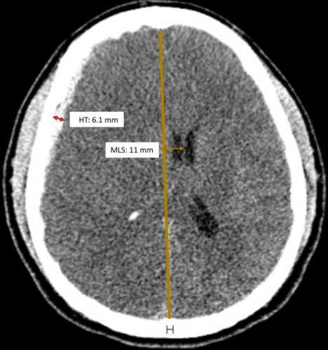
Source: Trauma Surgery Acute Care Open

Zumkeller and colleagues first described the use of the mathematical difference between these two values in prognosticating outcomes in severe TBI in 1996.

Zumkeller Index (ZI) = Midline shift (MDI) – Hematoma thickness (HT)

Intuitively, we’ve been using this all along. At some point, we recognized that if the degree of midline shift exceeds the hematoma thickness, it’s a bad sign. The easiest way to explain this is that there is injury to the brain that is causing swelling so the shift is greater than the size of the hematoma. 

The authors of a recent paper from Brazil decided to quantify the prognostic value of the ZI by doing a post-hoc analysis of a previously completed prospective study.  They limited their study to adult patients with an acute traumatic subdural hematoma confirmed by CT scan. It used data from the 4-year period from 2012-2015.

They compared demographics and outcomes in three cohorts of ZI:

  • Zero or negative ZI, meaning that the midline shift was less than the size of the hematoma
  • ZI from 0.1 mm to 3.0 mm
  • ZI > 3.0 mm

And here are the factoids:’

  • A total of 114 patients were studied, and the mechanism of injury was about 50:50 from motor vehicle crashes vs falls
  • About two thirds were classified as severe and the others were mild to moderate, based on GCS
  • Median initial GCS decreased from 6 in the low ZI group to 3 in the highest ZI group, implying that injuries were worse in the highest ZI group
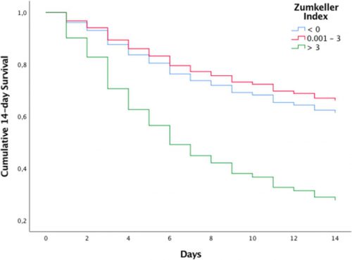
  • Mortality (14-day) was 91% in the highest ZI group and only in the low 30% range in the others
  • Regression analysis showed that patients with ZI > 3 had an 8x chance of dying within 14 days compared to the others
Source: Trauma Surgery Acute Care Open

Bottom line: This study confirms and quantifies something that many of us have been unconsciously using all along. Of course there are some possible confounding factors that were not quantified in this study. Patients with the more severe injuries tended to also have subarachnoid hemorrhage and/or intra-ventricular blood. Both are predictors of worse prognosis. But this is a nice study that quantifies our subjective impressions.

The Zumkeller Index is an easily applied tool using the measuring tool of your PACS application. It can be used to determine how aggressively to treat your patient, and may help the neurosurgeons decide who should receive a decompressive craniectomy and how soon.

So now go out and amaze your friends! You’ll be the life of the party!

Reference: Mismatch between midline shift and hematoma thickness as a prognostic factor of mortality in patients sustaining acute subdural hematomaTrauma Surgery & Acute Care Open 2021;6:e000707. doi: 10.1136/tsaco-2021-000707

Subdural Hematomas and Hygromas Simplified

There’s a lot of confusion about subdural pathology after head trauma. All subdural collections are located under the dura, on the brain’s surface. In some way, they involve or can involve the bridging veins, which are somewhat fragile and get more so with age.

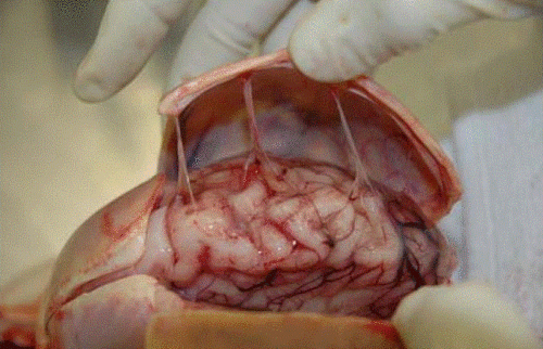
Head trauma causes a subdural hematoma by tearing some of these bridging veins. Notice how thick the dura is and how delicate the bridging veins are in the image below.

When these veins tear, bleeding ensues, which layers out over the surface of the brain in that area. If the bleeding does not stop, pressure builds and compresses and shifts the brain. A subdural hematoma is considered acute from the time of injury until about three days later. During this time, it appears more dense than brain tissue.

After about 3-7 days, the clot begins to liquefy and becomes less dense on CT. Many hematomas are reabsorbed, but occasionally there is repeated bleeding from the bridging veins, or the hematoma draws fluid into itself due to the concentration gradient. As a result, it can enlarge and begin to cause new symptoms. During this period, it is considered subacute.

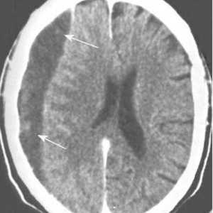
It moves on to a more chronic stage over the ensuing weeks. The blood cells in it break down completely, and the fluid that is left is generally less dense than the brain underneath it. The image below shows a chronic subdural (arrows).

Hygromas are different because they are a collection of CSF, not blood. They are caused by a tear in the meninges and allow CSF to accumulate in the subdural space. This can also be caused by head trauma and is generally very slow to form. They can lead to slow neurologic deterioration and are often found on head CT in patients with a history of falls, sometimes in the distant past. CT appearance is similar to a chronic subdural, but the density is the same as CSF, so it should have the same appearance as the fluid in the ventricle on CT.

What Is The Zumkeller Index in TBI?

I learned something new today: the Zumkeller index. Exciting! Most trauma professionals who take care of serious head trauma have already recognized the importance of quantifying extra-axial hematoma thickness (HT) and midline shift (MLS) of the brain. Here’s a picture to illustrate the concept:

Source: Trauma Surgery Acute Care Open

Zumkeller and colleagues first described the use of the mathematical difference between these two values in prognosticating outcomes in severe TBI in 1996.

Zumkeller Index (ZI) = Midline shift (MDI) – Hematoma thickness (HT)

Intuitively, we’ve been using this all along. At some point, we recognized that if the degree of midline shift exceeds the hematoma thickness, it’s a bad sign. The easiest way to explain this is that there is injury to the brain that is causing swelling so the shift is greater than the size of the hematoma. 

The authors of the current paper from Brazil decided to quantify the prognostic value of the ZI by doing a post-hoc analysis of a previously completed prospective study.  They limited their study to adult patients with an acute traumatic subdural hematoma confirmed by CT scan. It used data from the 4-year period from 2012-2015.

They compared demographics and outcomes in three cohorts of ZI:

  • Zero or negative ZI, meaning that the midline shift was less than the size of the hematoma
  • ZI from 0.1 mm to 3.0 mm
  • ZI > 3.0 mm

And here are the factoids:’

  • A total of 114 patients were studied, and the mechanism of injury was about 50:50 from motor vehicle crashes vs falls
  • About two thirds were classified as severe and the others were mild to moderate, based on GCS
  • Median initial GCS decreased from 6 in the low ZI group to 3 in the highest ZI group, implying that injuries were worse in the highest ZI group
  • Mortality (14-day) was 91% in the highest ZI group and only in the low 30% range in the others
  • Regression analysis showed that patients with ZI > 3 had an 8x chance of dying within 14 days compared to the others

Source: Trauma Surgery Acute Care Open

Bottom line: This study confirms and quantifies something that many of us have been unconsciously using all along. Of course there are some possible confounding factors that were not quantified in this study. Patients with the more severe injuries tended to also have subarachnoid hemorrhage and/or intra-ventricular blood. Both are predictors of worse prognosis. But this is a nice study that quantifies our subjective impressions.

The Zumkeller Index is an easily applied tool using the measuring tool of your PACS application. It can be used to determine how aggressively to treat your patient, and may help the neurosurgeons decide who should receive a decompressive craniectomy and how soon.

Reference: Mismatch between midline shift and hematoma thickness as a prognostic factor of mortality in patients sustaining acute subdural hematomaTrauma Surgery & Acute Care Open 2021;6:e000707. doi: 10.1136/tsaco-2021-000707
Delayed Intracranial Hemorrhage In Patients On Anticoagulants

A sizable portion of our population is taking one type of anticoagulant or another. Heck, even golf star Arnold Palmer and comedian Kevin Nealon are on Xarelto! Any trauma professional, and anyone who reads the package insert, knows that there is an increased risk of bleeding if they are injured while taking these drugs, whether it be warfarin or the new, novel anticoagulants.

But does the risk stop soon after injury? That is the presumption at many hospitals that initially treat these patients. They are seen in the ED, examined, scanned, and sent home if nothing is found. Is this a safe practice?

I have personally seen a patient who had an initially clean CT present within 12 hours after ED discharge with a catastrophic bleed and die. Yes, this is anecdotal, but I have talked to other trauma professionals with similar experiences. If this were just a minor complication, no big deal. But they died. Big problem for everyone involved.

So what does the literature say? Unfortunately, it consists of a collection of relatively small studies. Here are the collected factoids that I can glean from them:

  • Most are retrospective, observational studies 
  • Most are from a single hospital, which may miss readmissions to other facilities in the area
  • The delayed bleeding rate is about 0.5% to 1%
  • Some papers recommended discharging patients with a normal head CT and giving them instructions to return if new symptoms develop (this is what happened with my patient; what if they live alone or in a care center where these may not be recognized?!)
  • A few papers did identify patients needing neurosurgical intervention or who died
  • Immediate bleeds were more common with antiplatelet agents, delayed bleeds were more common with warfarin
  • I could find nothing that looked at this problem in patients taking novel anticoagulants like Pradaxa or Xarelto

Bottom line: The literature provides little guidance at this point. A good multi-institutional trial is needed to generate the numbers to tell us what to do. While we get around to this, I recommend that a selective brief observation (12 hrs) protocol be adopted. This protocol recognizes that subclinical bleeding may be present on initial presentation, and that a little more time is needed for it to declare itself.

Here is a link to our protocol. If the initial head CT is negative and the INR is less than 2.5, we will only discharge the patient if all of these criteria are true:

  • Age < 65
  • No skull fx
  • No new focal neurologic deficits
  • No soft tissue injury visible on CT (hematoma, laceration)
  • GCS = 15
  • No persistent vomiting
  • Brief TBI screen passed (Short Blessed Test, link here)

Most do not pass all of these, usually failing the age criterion. They are admitted for observation and neurologic monitoring for 12 hours, at which time the head CT is repeated. If it is still normal, then they can go home.

And although this protocol was designed with warfarin in mind, we apply it to patients taking novel anticoagulants like Pradaxa and Xarelto as well. We’ve had no epic fails yet, but I keep my fingers crossed!

Related posts:

References:

  • Management of minor head injury in patients receiving oral anticoagulant therapy: a prospective study of a 24-hour observation protocol. Ann Emerg Med 59(6):451-455, 2012.
  • Immediate and delayed traumatic intracranial hemorrhage in patients with head trauma and preinjury warfarin or clopidogrel use. Ann Emerg Med 59(6):460-468, 2012.
  • Delayed intracranial hemorrhage after blunt trauma: are patients on preinjury anticoagulants and prescription antiplatelet agents at risk? J Trauma 71(6):1600-1604, 2011.
  • Low risk of late intracranial complications in mild traumatic brain injury patients using oral anticoagulation after an initial normal brain computed tomography scan: education instead of hospitalization. Eur J Neurol 21(7):1021-1025, 2014.
  • Can anticoagulated patients be discharged home safely from the emergency department after minor head injury? J Emerg Med 46(3):410-417, 2014.
  • Patients with blunt head trauma on anticoagulation and antiplatelet medications: can they be safely discharged after a normal initial cranial computed tomography scan? Am Surg 80(6):610-613, 2014.

Subdural Hematomas and Hygromas Simplified

There’s a lot of confusion about subdural pathology after head trauma. All subdural collections are located under the dura, on the surface of the brain. In some way they involve or can involve the bridging veins, which are somewhat fragile and get more so with age.

Head trauma causes a subdural hematoma by tearing some of these bridging veins. Notice how thick the dura is and how delicate the bridging veins are in the image below.

When these veins tear, bleeding ensues which layers out over the surface of the brain in that area. If the bleeding does not stop, pressure builds and begins compressing and shifting the brain. A subdural hematoma is considered acute from time of injury until about 3 days later. During this time, it appears more dense than brain tissue.

After about 3-7 days, the clot begins to liquefy and becomes less dense on CT. Many hematomas are reabsorbed, but occasionally there is repeated bleeding from the bridging veins, or the hematoma draws fluid into itself due to the concentration gradient. It can enlarge and begin to cause new symptoms. During this period it is considered subacute.

It moves on to a more chronic stage over the ensuing weeks. The blood cells in it break down completely, and the fluid that is left is generally less dense than the brain underneath it. The image below shows a chronic subdural (arrows).

Hygromas are different, in that they are a collection of CSF and not blood. They are caused by a tear in the meninges and allow CSF to accumulate in the subdural space. This can be caused by head trauma as well, and is generally very slow to form. They can lead to slow neurologic deterioration, and are often found on head CT in patients with a history of falls, sometimes in the distant past. CT appearance is similar to a chronic subdural, but the density is the same as CSF, so it should have the same appearance as the fluid in the ventricle on CT.

Related posts: