Tag Archives: fracture

What You Need To Know About: Frontal Sinus Fractures

Fracture of the frontal sinus is less common than other facial injuries, but can be more complex to deal with, both in the shorter and longer terms. These are generally high energy injuries, and facial impact in car crashes is the most common mechanism. Fists generally can’t cause the injury, but blunt objects like baseball bats can.

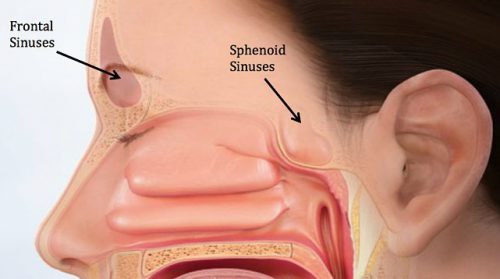
Here’s the normal anatomy:

sinus-fracture-treatment


Source: www.facialtraumamd.com

There are two “tables”, the anterior and the posterior. The anterior is covered with skin and a small amount of subcutaneous tissue. The posterior table is separated from the brain by the meninges.

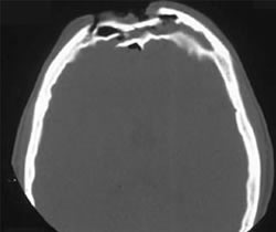
Here’s an image of an open fracture involving both tables. Note the underlying pneumocephalus.

frontal_sinus1

A third of injuries violate the anterior table, and two thirds violate both. Posterior table fractures are very rare. A third of all patients will develop a CSF leak, typically from their nose.

These fractures may be (rarely) identified on physical exam if deformity and flattening is noted over the forehead. Most of the time, these patients undergo imaging for brain injury and the fracture is found incidentally. Once identified, go back and specifically look for a CSF leak. Clear fluid in the nose is, by definition, CSF. Don’t waste time on a beta-2 transferring (see below).

If a laceration is clearly visible over the fracture, or if a CSF leak was identified, notify your maxillofacial specialist immediately. If more than a little pneumocephalus is present, let your neurosurgeon know. Otherwise, your consults can wait until the next morning.

In general, these patients frequently require surgery for the fracture, either to restore cosmetic contours or to avoid mucocele formation. However, these are seldom needed urgently unless the fracture is an open fracture with contamination or there is a significant CSF leak. If in doubt, though, consult your specialist.

Related posts:

NSAIDs And Fracture Healing Revisited

Over the years, I’ve commented several times on the “myth” of NSAIDs causing problems with fracture healing. I still hear occasional comments from my orthopedic colleagues cautioning against the use of these drugs in patients who have had fracture repairs.

But is it true?  In 2003, several papers brought to light possible interactions between these drugs and fracture healing. Specifically, there were questions about these drugs interfering with the healing process and of increasing the number of delayed unions or nonunions. But once again, how convincing were these papers, really?

It would seem to make sense that NSAIDs could interfere with bone healing. The healing process relies heavily on the regulation of osteoblast and osteoclast function, which itself is regulated by prostaglandins. Since prostaglandins are synthesized by the COX enzymes, COX inhibitors like the NSAIDs should have the potential to impair this process. Indeed, animal studies in rats and rabbits seem to bear this out.

But as we have seen before, good animal studies don’t always translate well into human experience. Although a study from 2005 suggested that NSAID administration in older patients within 90 days of injury had a higher incidence of fracture nonunion, the study design was not a very good one. It was equally likely that patients who required these drugs in this age group may have been at higher risk for nonunion in the first place.

A meta-analysis of human studies was performed in 2011. Out of 558 potential studies, only 5 met criteria review. (This is yet another reminder of the sheer amount of sub-par research out there.) The authors found that short-term use (< 14 days) of normal dose NSAIDS was not associated with non-union. High doses of ketorolac (> 120mg/day) and diclofenac sodium (> 300mg total) did have an association. But remember, this does not show causation. There are many other factors that can impede healing (smoking, diabetes, etc).

A study from 2016 examined the effect of ketorolac administration on fracture healing in patients undergoing repairs of femoral and tibial fractures. It did not find an association between non-union and ketorolac, but did find one with smoking. Unfortunately, the study was small (85 patients given ketorolac, 243 controls without it). It probably does not have the statistical power to detect any difference with the NSAID. A power analysis was not provided in the methods section.

Bottom line: Once again, the animal data is clear and the human data less so. Although there are theoretical concerns about NSAID use and fracture healing, there is still not enough solid risk:benefit information to abandon short-term NSAID use in patients who really need them. NSAIDs can and should be prescribed in patients with short-term needs and simple fractures. But we now have evidence that high-dose NSAIDs may have some impact, so stick to the usual doses for just as long as they are needed for pain management.

References:

  1. Effects of nonsteroidal anti-inflammatory drugs on bone formation and soft-tissue healing. J AM Acad Orthop Surg 12:139-43, 2004.
  2. Effect of COX-2 on fracture-healing in the rat femur. J Bone Joint Surg Am 86:116-123, 2004.
  3. Effects of perioperative anti-inflammatory and immunomodulating therapy on surgical wound healing. Pharmacotherapy 25:1566-1591, 2005.
  4. Pharmacological agents and impairment of fracture healing: what is the evidence? Injury 39:384-394, 2008.
  5. High dose nonsteroidal anti-inflammatory drugs compromise spinal fusion. Can J Anaesth 52:506-512, 2005.
  6. Nonsteroidal Anti-Inflammatory Drugs and Bone-Healing: A Systematic Review of Research Quality. JBJS Rev 4(3), 2016.
  7. High-dose ketorolac affects adult spinal fusion. Spine 36(7):E461-E468, 2011.
  8. Ketorolac administered in the recovery room for acute pain management does not affect healing rates of femoral and tibial fractures. J Orthop Trauma 30(9):479-482, 2016.
What Is: A Pars Fracture / Defect

Radiologists sure know their anatomy! The vast majority of the time, I actually know what they are describing. But every once in a while they’ll toss in some term that I know I probably learned about in medical school (last century). For whatever reason though, I’m just not able to retrieve it.

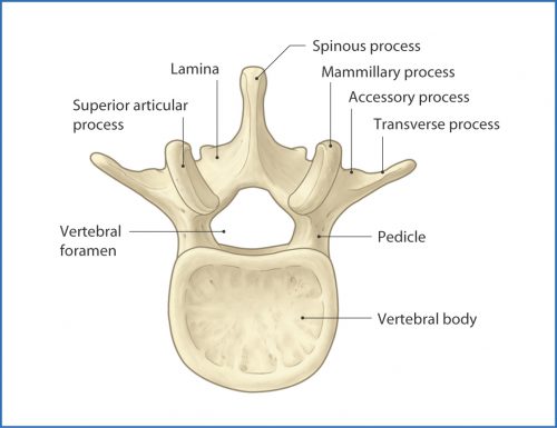
Which brings me to the pars fracture. Hmm. I figure that if I have to hit the books again to look something up, there are probably a few other trauma professionals out there who are dying to know what it is, too. Here’s a diagram of a typical vertebra:

The arch extending away from the vertebral body consists of the pedicles, which are connected by the lamina. A number of things jut off from this arch, including the transverse and spinous processes and the articular processes.

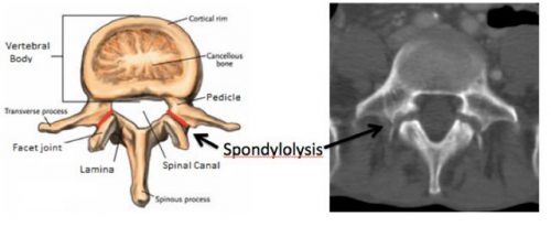
The area between the lamina and pedicle and adjacent to the articular process is called the pars interarticularis. This area is a bit thinner and flatter than the rest of the arch and can fracture if sufficient acute stress is applied. It can also fracture if enough chronic stress in the area occurs. This pattern is typically seen in the lumbar spine, but may also occur at the cervical level. Thus, a pars fracture or pars defect is simply a fracture through this area.

Another term you may see with regard to the pars is spondylolysis. This is defined as a defect in the pars interarticularis, typically from a fracture. So if you see either of these terms in a radiology report, recognize that they are basically one and the same.

Here is a nice image showing the location of the pars, and the axial CT appearance of “bilateral pars defects.”

Mystery solved! Amaze your friends!

Giving Vitamin D After Fracture

The role of Vitamin D in fracture healing is well known. So, of course, trauma professionals have tried to promote Vitamin D

supplementation to counteract the effects of osteoporosis. A meta-analysis of of 12 papers on the topic relating to hip and other non-vertebral fractures showed that there was roughly a 25% risk reduction for any non-vertebral fractures in patients taking 700-800 U of Vitamin D supplements daily.

Sounds good. So what about taking Vitamin D after a fracture occurs? Seems like it should promote healing, right? A very recent meta-analysis that is awaiting publication looked at this very question.

Unfortunately, there was a tremendous variability in the interventions, outcomes, and measures of variance. All the authors could do was summarize individual papers, and a true meta-analysis could not be performed.

Here are the factoids:

  •  81 papers made the cut for final review
  • A whopping 70% of the population with fractures had low Vitamin D levels
  • Vitamin D supplementation in hospital and after discharge did increase serum levels
  • Only one study, a meeting abstract which has still not seen the light of day in a journal, suggested a trend toward less malunions following a single loading dose of Vitamin D

Bottom line: Vitamin D is a great idea for people who are known to have, or are at risk for, osteoporosis and fractures. It definitely toughens up the bones and lowers the risk of fracture. However, the utility of giving it after a fall has not been shown. Of the 81 papers reviewed, none showed a significant impact on fracture healing. The only good thing is that Vitamin D supplements are cheap. Giving them may make us think that we are helping our patient heal, but it’s not. 

References:  

  • What is the role of vitamin D supplementation in acute fracture patients? A systematic review and meta-analysis of the prevalence of hypovitaminosis D and supplementation efficacy. J Orthopaedic Trauma epub Sep 22 2015.
  • Fracture prevention with vitamin D supplementation: a meta-analysis of randomized controlled trials. JAMA 293(18):2257-2264, 2005.

What Is: The Monteggia Fracture

Yesterday, I wrote about one of the many fractures that occurs during falls onto outstretched hands, the Galeazzi fracture. Today, I’ll describe another one, the Monteggia fracture. Yes, this one is named after another Italian surgeon! And like the other one, the person it was named after was actually the second to describe it.

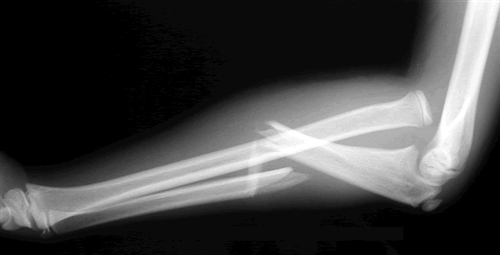
Think of the Monteggia fracture as the exact opposite of a Galeazzi fracture. The fractured bone is switched, as is the dislocation. Whereas the Galeazzi is a distal radius fracture with a distal ulnar dislocation which pulls the radio-ulnar joint apart, the Monteggia is a proximal ulnar fracture with a proximal radial head dislocation.

Here’s what it looks like:

Of course, the orthopedic surgeons have a classification system for this based on the directions the bones fracture and dislocate. I won’t bore you with the details.

Unlike the Galeazzi fracture, all of these require operative repair, even in children. This helps stabilize the radial head and decreases the incidence of malunion.