Category Archives: Imaging

Arms Up or Arms Down In Torso CT Scans?

CT scan is a valuable tool for initial screening and diagnosis of trauma patients. However, more attention is being paid to radiation exposure and dosing. Besides selecting patients carefully and striving for ALARA radiation dosing (as low as reasonably achievable) by adjusting technique, what else can be done? Obviously, shielding parts of the body that do not need imaging is simple and effective. But what about simply changing body position?

One simple item to consider is arm positioning in torso scanning. There are no consistent recommendations for use in trauma scanning. Patients with arm and shoulder injuries generally keep the affected upper extremity at their side. Radiologists prefer to have the arms up if possible to reduce scatter and provide clearer imaging.

Radiation physics research has examined arm positioning and its effect on radiation dose. A retrospective review of 690 patients used dose information computed by the CT software and displayed on the console. Radiation exposure was estimated using this data and was stratified by arm positioning. Even though there are some issues with study design, the results were impressive.

The dose results were as follows:

  • Both arms up: 19.2 mSv (p<0.0000001)
  • Left arm up: 22.5 mSv
  • Right arm up: 23.5 mSv
  • Arms down: 24.7 mSv

Bottom line: Do everything you can to reduce radiation exposure:

  1. Be selective with your imaging. Do you really need it?
  2. Work with your radiologists and physicists to use techniques that reduce dose yet retain image quality
  3. Shield everything that’s not being imaged.
  4. Think hard about getting CT scans in children
  5. Raise both arms up during torso scanning unless injuries preclude it.

Reference: Influence of arm positioning on radiation dose for whole body computed tomography in trauma patients. J Trauma 70(4):900-905, 2011.

The Role Of Postop CT Scan In Penetrating Trauma

CT scans are commonly used to aid the workup of patients with blunt trauma. They are occasionally useful in penetrating trauma, specifically when penetration into a body cavity is uncertain, and the patient has no hard signs that would send him or her immediately to the operating room.

Is there any role for CT in operative penetrating trauma, after the patient has already been to the OR? The dogma has always been that the eyeballs of the surgeon in the OR are better than any other imaging modality. Really? The surgical group at San Francisco General addressed this question by retrospectively reviewing 6 years of their operative penetrating injury registry data. They were interested in finding how many occult injuries (seen with CT but not by the surgeon) were found on a postop CT. A total of 225 patients who underwent operative management of penetrating abdomen or chest injury were included. Here are the factoids:

  • Only 110 patients had a postop CT scan; 73 had scans within the first 24 hours, the other 37 were scanned later
  • The rationale for early scan was to investigate retroperitoneal injury in half of patients, but frequently no indication was given (41%)
  • The rationale for late scan was for workup of ileus in one-third or for evaluation of new or unexpected clinical problems
  • Occult injuries were found in about half of early CT patients (52%) and 22% of late CT patients
  • The most common occult injuries were fractures, GU issues, regraded solid organ injury, and unrecognized vascular injuries
  • Ten patients had management changes, including:
    • Interventional radiology for four injuries with extravasation
    • Operation for orthopedic or GU injury in seven patients
    • One patient underwent surgery for an unstable spine fracture

Bottom line: There appears to be a significant benefit to sending some penetrating injury patients to CT in the early postop period. Specifically, those with injury to the retroperitoneum, deep into the liver, near the spine, or with multiple and complicated injuries would benefit. Simple stabs and gunshots that stay away from these areas/structures probably do not need follow-up imaging. 

Reference: Routine computed tomography after recent operative exploration for penetrating trauma: What injuries do we miss? J Trauma 83(4):575-578, 2017.

Updated: How To Detect Bucket Handle Injuries With CT

A bucket-handle injury is a relatively uncommon complication of blunt trauma to the abdomen. It only occurs in a few percent of patients, but is much more likely if they have a seat belt sign.  The basic pathology is that the bowel mesentery (small bowel of sigmoid colon) gets pulled away from the intestinal wall.

This injury is problematic because it may take a few days for the bowel itself to die and perforate. Patients with no other injuries could potentially be discharged from the hospital before they become overtly symptomatic, leading to delayed treatment.

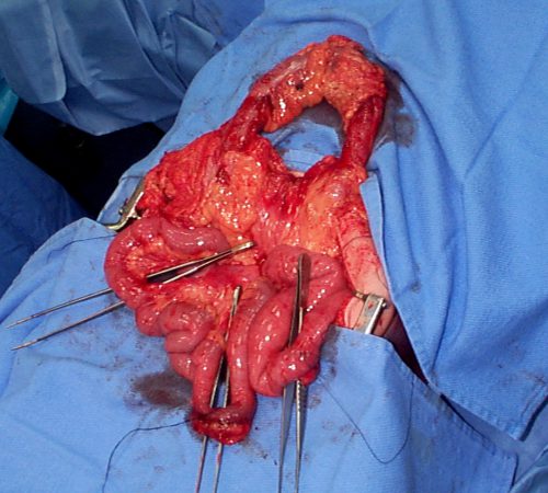
Here’s an image from my personal collection with not one, but four bucket-handle injuries.

Typical patients with suspected blunt intestinal injury are observed with good serial exams and a daily WBC count. If this begins to rise after 24 hours, there is a reasonable chance that this injury is present.

CT scan has not really been that reliable in past studies. There may be some “dirty mesentery”, which is contused and has a hematoma within it. But without a more convincing exam, it is difficult to convince yourself to operate immediately on these patients.

A paper was published by a group of radiologists at Duke University. It appears to be a case report disguised as a descriptive paper. It looks like they picked a few known bucket-handle injuries from their institution and back-correlated them with CT findings.

The authors called out the usual culprits:

  • Fluid between loops of bowel
  • Active bleeding in the mesentery
  • Bowel wall perfusion defects

But they also noted that traumatic abdominal wall hernias were highly associated with seat belt sign as well. These are rare, but should bring intestinal injury to mind when seen.

With newer scanners, radiologists are better able to detect subtle areas of hypoperfusion as well. This is a fairly good indicator of injury, especially when adjacent bowel appears normally perfused. Here are two examples. The black arrows denote active extravasation, and the white ones an area of hypoperfusion.

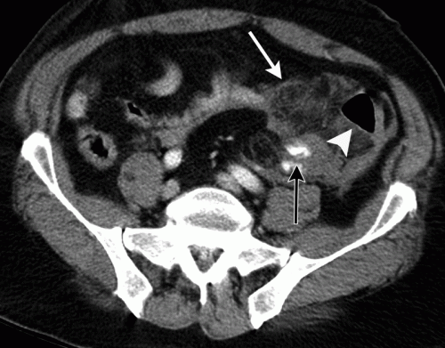
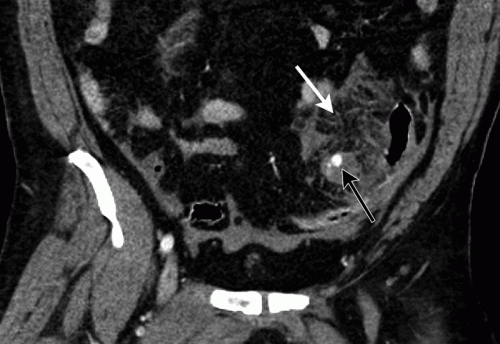
The authors add bowel wall hypoperfusion as another finding that may point to a bucket-handle type injury.

A recent paper demonstrates the value of the current generation of high-quality scanners. A collection of California and Denver centers implemented a multicenter, prospective, observational study of patients with seat belt signs. The developed a list of positive findings, which included:

  • abdominal wall soft tissue contusion (radiographic seat belt sign)
  • free peritoneal fluid
  • bowel wall thickening
  • mesenteric stranding
  • mesenteric hematoma
  • bowel dilation
  • pneumatosis
  • pneumoperitoneum

A total of 754 patients with visible seat belt sign were enrolled and all went to CT scan. Any of the findings listed above were associated with a statistically significant likelihood of hollow viscus injury. The highest likelihood was associated with:

  • free peritoneal fluid – 42x more likely
  • bowel dilation – 21x
  • free fluid with no solid organ injury – 20x
  • bowel wall thickening – 19x
  • radiographic seat belt sign – 3x

Any of the radiographic findings strongly suggested that an injury could be present. However, if none were present, it was very unlikely that there were any significant injuries. The authors suggested that if such patients had no other injuries requiring hospitalization, they could potentially be discharged home. However, those patients should be counseled to return for evaluation immediately if they have any change in their abdominal or systemic status.

Bottom line: Some patients with a visible seat belt sign might be eligible for discharge from the ED if they have a totally negative abdominal CT and no other injuries requiring hospitalization. If they have any finding, they should be admitted for observation.

If your patient has an unconcerning exam and any of the findings listed above, perform serial exams and get a WBC the next morning. If the exam worsens, operate. If the WBC rises, consider laparoscopy to see if you need to make a bigger incision. And if you see any evidence of hypoperfused bowel, consider laparoscopy right away. 

References:

  • Excluding Hollow Viscus Injury for Abdominal Seat Belt Sign Using Computed Tomography. JAMA Surg. 2022 Sep 1;157(9):771-778. doi: 10.1001/jamasurg.2022.2770. PMID: 35830194; PMCID: PMC9280606.
  • CT findings of traumatic bucket-handle mesenteric injuries. Am J Radiol 209:W360-@364, 2017.
  • Multidetector CT of blunt abdominal trauma. Radiology 265(3):678–693, 2012.

More On MRI And External Fixators

I’ve covered the problem of performing MRI on patients with external fixators. This is typically a problem that arises in head-injured patients with extremity or pelvic fixators for concomitant fractures.

MRI is an indispensable tool for the evaluation of head, spine, and soft tissue trauma. However, a great deal of effort is required to ensure that any patient scheduled for this test is “MRI compatible.” The fear is that any retained metallic fragments may move or heat up once the magnets are activated.

But what about trauma patients with external fixators? That is one big hunk of metal inserted deep into your patient. There are three major concerns:

  • Is the material ferromagnetic? If so, it will move when the magnets are activated and may cause internal injury. These days, many fixator sets are not ferromagnetic, avoiding this problem.
  • Can currents be induced in the material, causing heating? This is not much of a problem for small, isolated objects. However, external fixators are configured so that current loops can be created. The fluctuating magnetic fields can induce currents that, in turn, will heat the surrounding tissue. And thinner materials (narrow pins) result in more current and heating.
  • Will the metal degrade image quality?

Thankfully, there is a continuing trickle of evidence that is accumulating to give us some guidance. One paper from 2017 described a retrospective case series from four trauma centers. The authors performed MRIs on 38 patients with 44 external fixators. The adverse events they monitored for were catastrophic hardware pullout, thermal injury to the skin, field distortions that impaired the images, and damage to the magnet casing.

Twelve patients with 13 external fixators had MIR performed with the hardware inside the MRI bore, and 27 patients had the study with the fixator outside the bore. Most MRIs were performed to evaluate the cervical spine. There were no adverse events.

A recent Massachusetts General Hospital study involved a larger group (97 patients with 110 fixators). The fixators were located on the ankles, knee, femur, and pelvis. Most were performed on a 1.5T MRI, although a few were done on a 3T machine. Again, most scans were performed for head or cervical spine evaluation. Two of the 97 studies were terminated early due to patient discomfort. In both cases, the frame was outside the MRI bore.

The biggest challenge in our clinical practice is that there is no standard ex-fix configuration. Our orthopedic colleagues get to unleash their creativity, trying to devise the appropriate architecture to hold bones together so they can heal properly. This makes developing standardized guidelines regarding what can and can’t go into the scanner difficult.

We do know from clinical simulation studies that heating is influenced by ex-fix configuration. Increasing pin depth (thicker extremities) and closer pin spacing produces smaller temperature rises. For example, pins placed in a 15cm bar at a depth of 11cm produced a temperature rise of 2 degrees, but pins placed along a 30cm bar at a depth of 2cm showed a rise of 6 degrees.

However, a growing body of literature shows that the heating effects are relatively small and get smaller as the distance from the magnet increases. And non-ferromagnetic materials move very little, if at all, and do not interfere with the image. So as long as nonferromagnetic materials are used, the patients are probably safe as long as basic principles are adhered to:

  • Other diagnostic options should be considered and/or exhausted prior to using MRI.
  • Informed consent must be obtained, explaining that the potential risks are not completely understood.
  • The fixator must be tested with a handheld magnet so that all ferromagnetic components can be identified and removed.
  • All traction bows must be removed.
  • Ice bags or cooling packs should be placed at all skin-pin interfaces.
  • The external fixator should remain at least 7cm outside the bore at all times, if possible. If any portion must be inside the bore, monitoring efforts should be stepped up even more.

Bottom line: MRI of patients with external fixators can be safely accomplished. Consult your radiologists and physicists to develop a policy that is specific to the scanners used at your hospital. 

References:

  1. Magnetic Resonance Imaging of Trauma Patients Treated With Contemporary External Fixation Devices: A Multicenter Case Series. Journal of Orthopaedic Trauma, 31 (11), e375-e380. doi: 10.1097/BOT.0000000000000954.
  2. Magnetic Resonance Imaging of Trauma Patients Treated With Contemporary External Fixation Devices: A Multicenter Case Series. J Orthop Trauma. 2017 Nov;31(11):e375-e380. doi: 10.1097/BOT.0000000000000954. PMID: 28827510.


New Technology: Using AI To Interpret Pelvic X-rays

Look out, radiologists! The computers are coming for you!

Radiologists use their extensive understanding of human anatomy and combine it with subtle findings they see on x-ray shadow pictures. In doing this, they can identify a wide variety of diseases, anomalies, and injuries. But as we have seen with vision systems and game playing (think chess), computers are getting pretty good at doing this as well.

Is it only a matter of time until computer artificial intelligence (AI) starts reading x-rays?  Look at how good they already are at interpreting EKGs. The trauma group at Stanford paired up with the Chang Gung Memorial Hospital in Taiwan to test the use of AI for interpreting images to identify a specific set of common pelvic fractures.

The Stanford group used a deep learning neural network (XCeption) to analyze source x-rays (standard A-P pelvis images) from Chang Gung. These x-rays were divided into training and testing cohorts. The authors also applied different degrees of blurring, brightness, rotation, and contrast adjustment to the training set in order to help the AI overcome these issues when interpreting novel images.

The AI interpreted the test images with a very high degree of sensitivity, specificity, accuracy, and predictive values, with all of them over 0.90. The algorithms generated a “heat map” that showed the areas that were suspicious for fracture. Here are some examples with the original x-ray on the left and the heat map on the right:

The top row shows a femoral neck fracture, the middle row an intertrochanteric fracture, and the bottom row another femoral neck fracture with a contralateral implant. All were handily identified by the AI.

AI applications are usually only as good as their training sets. In general, the bigger the better so they can gain a broader experience for more accurate interpretation. So it is possible that uncommon, subtle fractures could be missed. But remember, artificial intelligence is meant to supplement the radiologist, not replace him or her. You can all breathe more easily now.

This technology has the potential for broader use in radiographic interpretation. In my mind, the best way to use it is to first let the radiologist read the images as they usually do. Once they have done this, then turn on the heat map so they can see any additional anomalies the AI has found. They can then use this information to supplement the initial interpretation.

Expect to see more work like this in the future. I predict that, ultimately, the picture archiving and communications systems (PACS) software providers will build this into their product. As the digital images are moving from the imaging hardware to the digital storage media, the AI can intercept it and begin the augmented interpretation process. The radiologist will then be able to turn on the heat map as soon as the images arrive on their workstation.

Stay tuned! I’m sure there is more like this to come!

Reference: Practical computer vision application to detect hip fractures on pelvic X-rays: a bi-institutional study.  Trauma Surgery and Acute Care Open 6(1), http://dx.doi.org/10.1136/tsaco-2021-000705.