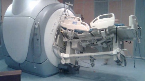
I’ve covered the problem of performing MRI on patients with external fixators. This is typically a problem that arises in head-injured patients with extremity or pelvic fixators for concomitant fractures.
MRI is an indispensable tool for the evaluation of head, spine, and soft tissue trauma. However, a great deal of effort is required to ensure that any patient scheduled for this test is “MRI compatible.” The fear is that any retained metallic fragments may move or heat up once the magnets are activated.

But what about trauma patients with external fixators? That is one big hunk of metal inserted deep into your patient. There are three major concerns:
- Is the material ferromagnetic? If so, it will move when the magnets are activated and may cause internal injury. These days, many fixator sets are not ferromagnetic, avoiding this problem.
- Can currents be induced in the material, causing heating? This is not much of a problem for small, isolated objects. However, external fixators are configured so that current loops can be created. The fluctuating magnetic fields can induce currents that, in turn, will heat the surrounding tissue. And thinner materials (narrow pins) result in more current and heating.
- Will the metal degrade image quality?
Thankfully, there is a continuing trickle of evidence that is accumulating to give us some guidance. One paper from 2017 described a retrospective case series from four trauma centers. The authors performed MRIs on 38 patients with 44 external fixators. The adverse events they monitored for were catastrophic hardware pullout, thermal injury to the skin, field distortions that impaired the images, and damage to the magnet casing.
Twelve patients with 13 external fixators had MIR performed with the hardware inside the MRI bore, and 27 patients had the study with the fixator outside the bore. Most MRIs were performed to evaluate the cervical spine. There were no adverse events.
A recent Massachusetts General Hospital study involved a larger group (97 patients with 110 fixators). The fixators were located on the ankles, knee, femur, and pelvis. Most were performed on a 1.5T MRI, although a few were done on a 3T machine. Again, most scans were performed for head or cervical spine evaluation. Two of the 97 studies were terminated early due to patient discomfort. In both cases, the frame was outside the MRI bore.
The biggest challenge in our clinical practice is that there is no standard ex-fix configuration. Our orthopedic colleagues get to unleash their creativity, trying to devise the appropriate architecture to hold bones together so they can heal properly. This makes developing standardized guidelines regarding what can and can’t go into the scanner difficult.
We do know from clinical simulation studies that heating is influenced by ex-fix configuration. Increasing pin depth (thicker extremities) and closer pin spacing produces smaller temperature rises. For example, pins placed in a 15cm bar at a depth of 11cm produced a temperature rise of 2 degrees, but pins placed along a 30cm bar at a depth of 2cm showed a rise of 6 degrees.
However, a growing body of literature shows that the heating effects are relatively small and get smaller as the distance from the magnet increases. And non-ferromagnetic materials move very little, if at all, and do not interfere with the image. So as long as nonferromagnetic materials are used, the patients are probably safe as long as basic principles are adhered to:
- Other diagnostic options should be considered and/or exhausted prior to using MRI.
- Informed consent must be obtained, explaining that the potential risks are not completely understood.
- The fixator must be tested with a handheld magnet so that all ferromagnetic components can be identified and removed.
- All traction bows must be removed.
- Ice bags or cooling packs should be placed at all skin-pin interfaces.
- The external fixator should remain at least 7cm outside the bore at all times, if possible. If any portion must be inside the bore, monitoring efforts should be stepped up even more.
Bottom line: MRI of patients with external fixators can be safely accomplished. Consult your radiologists and physicists to develop a policy that is specific to the scanners used at your hospital.
References:
- Magnetic Resonance Imaging of Trauma Patients Treated With Contemporary External Fixation Devices: A Multicenter Case Series. Journal of Orthopaedic Trauma, 31 (11), e375-e380. doi: 10.1097/BOT.0000000000000954.
- Magnetic Resonance Imaging of Trauma Patients Treated With Contemporary External Fixation Devices: A Multicenter Case Series. J Orthop Trauma. 2017 Nov;31(11):e375-e380. doi: 10.1097/BOT.0000000000000954. PMID: 28827510.

