Tag Archives: imaging

How Much Fetal Radiation Exposure In Imaging Studies?

I periodically publish a chart that shows how much radiation exposure our patients get from various trauma imaging studies. For reference, here it is:

Test Dose (mSv) Equivalent background
radiation
Chest x-ray 0.1 10 days
Pelvis x-ray 0.1 10 days
CT head 2 8 months
CT cervical spine 3 1 year
Plain c-spine 0.2 3 weeks
CT chest 7 2 years
CT abdomen/pelvis 10 3 years
CT T&L spine 7 2 years
Plain T&L spine 3 1 year
Millimeter wave
scanner (that hands
in the air TSA thing at
the airport)
0.0001 15 minutes
Scatter from a chest
x-ray in trauma bay
when standing one 
meter from the
patient
0.0002 45 minutes
Scatter from a chest
x-ray in trauma bay
when standing three 
meters from the
patient
0.000022 6 minutes

One of the issues that trauma professionals gnash our teeth about is how much radiation the baby gets when we perform these studies on pregnant women. Well, here is just what you need. Another chart! In order to avoid confusion, I will list effective doses to the fetus in milligrays (mGy), which is how much radiation is deposited in a substance. This is a little confusing, since doses are frequently listed in millisieverts which takes the specific organ and type of radiation into effect. In general, these two units are very similar for x-rays.

A useful rule of thumb is that if the fetal dose is less than 50 mGy during any trimester, the risk of an abortion or fetal malformation is about the same as from other risks to the pregnancy. The American College of Radiology notes that exposures less than 100 mGy are “probably too subtle to be clinically detectable.”

To help in your clinical decision making, I’ve added some extra information to the table regarding fetal exposure:

Test Adult Dose (mSv) Fetal Dose (mGy)
Chest x-ray 0.1 negligible
Pelvis x-ray 0.1 negligible
CT head 2 <1
CT head and C-spine 4 10
CT chest 7 <1
CT abdomen/pelvis 10 25
Pan Scan (CTA chest, abdomen, and pelvis) up to 68 up to 56
CT pulmonary angiogram up to 40 <1

Bottom line: We still have to think hard about how we image pregnant patients! There are some alternatives available to us, including the good old physical exam, conventional x-rays, and ultrasound. MRI is possible, but is a pain in the ass for many reasons. 

CT of the head and cervical spine are fine for both mother and baby, and non-contrast imaging of the torso is within accepted limits of fetal exposure. However, the whole point of the torso scan in CT is to identify critical injuries that may lead to exsanguination like solid organ and aortic injuries. In general, those scans should always be ordered with contrast. 

If clinical suspicion is high, it may be necessary to order these higher-dose studies anyway. If the mother has an unrecognized and potentially fatal injury, the baby will not survive either. There are many, many permutations of injuries and diagnostics. These cases will put your clinical judgment to the test, for sure!

References:

  • Imaging Pregnant and Lactating patients. RadioGraphics 35:6, 1751-1765, 2015.
  • Imaging of the pregnant trauma patient. RadioGraphics 34:3, 748-763,2014.
  • Fetal doses from radio logical examinations. Br J Radiol;72(860): 773–780, 1999.

Best Of EAST 2023 #8: Use Of AI To Detect Rib Fractures On CT

Artificial intelligence systems (AI) are increasingly finding their way into medical practice. They have been used to assist pathologists in screening microscope specimens for years. Although still amazingly complicated, one of the most obvious applications for trauma is in reading x-rays. Counting rib fractures may be helpful for care planning, and characterizing fracture patterns may assist our orthopedic colleagues in evaluating and planning rib plating procedures.

The trauma group at Stanford developed a computer vision system to assist in identifying fractures and their percent displacement.  They used a variation on a neural network deep learning system and trained it on a publicly available CT scan dataset.  They used an index of radiographic similarity (DICE score) to test how well their model matched up against the reading of an actual radiologist.

Here are the factoids:

  • The AI network was trained on a dataset of 5,000 images in 660 chest CT scans that had been annotated by radiologists
  • The model achieved a DICE score of 0.88 after training
  • With a little jiggering of the model (reweighting), the receiver operating characteristic curve improved to 0.99, which is nearly perfect

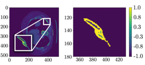
The left side shows a CT scan rotated 90 degrees; the right side shows the processed data after a fracture was detected.

Bottom line: This paper describes what lies ahead for healthcare in general. The increasing sophistication and accuracy of AI applications will assist trauma professionals in doing their jobs better. But rest easy, they will not take our jobs anytime soon. What we do (for the most part) takes very complex processing and decision making. It will be quite some time before these systems can do anything more that augment what we do.

Expect to see these AI products integrated with PACS viewing systems at some point in the not so distant future. The radiologist will interpret images in conjunction with the AI, which will highlight suspicious areas on the images as an assist. The radiologist can then make sure they have reported on all regions that both they and the AI have flagged.

Here are my questions and comments for the presenter/authors:

  • How can you be sure that your model isn’t only good for analyzing your training and test datasets? If neural networks are overtrained, they get very good at the original datasets but are not so good analyzing novel datasets. Have you tried the on your own data yet?
  • Explain what “class reweighting” is and how it improved your model. I presume you used this technique to compensate for the potential issue mentioned above. But be sure to explain this in simple terms to the audience.
  • Don’t lose the audience with the net details. You will need to give a basic description of how deep learning nets are developed and how they work, but not get too fancy.

This is an interesting glimpse into what is coming to a theater near you, so to speak. Expect to see applications appearing in the next few years.

Reference: AUTOMATED RIB FRACTURE DETECTION AND CHARACTERIZATION ON COMPUTED TOMOGRAPHY SCANS USING COMPUTER VISION. EAST 2023 Podium paper #16.

Best Of EAST 2023 #5: Imaging The Elderly

Several papers have been published over the years regarding underdiagnosis when applying the usual imaging guidelines to elderly trauma patients. Unfortunately, our elders are more fragile than the younger patients those guidelines were based on, leading to injury from lesser mechanisms. They also do not experience pain the same way and may sustain serious injuries that produce no discomfort on physical exam. Yet many trauma professionals continue to apply standard imaging guidelines that may not apply to older patients.

EAST sponsored a multicenter trial on the use of CT scans to minimize missed injuries. Eighteen Level I and Level II trauma centers prospectively enrolled elderly (age 65+) trauma patients in the study over one year. Besides the usual demographic information, data on physical exams, imaging studies, and injuries identified were also collected. The study sought to determine the incidence of delayed injury diagnosis, defined as any identified injury that was not initially imaged with a CT scan.

Here are the factoids:

  • Over 5,000 patients were enrolled, with a median age of 79
  • Falls were common, with 65% of patients presenting after one
  • Nearly 80% of patients actually sustained an injury (!)
  • Head and cervical spine were imaged in about 90% of patients, making them the most common initial studies
  • The most commonly missed injuries involved BCVI (blunt carotid and vertebral injury) or thoracic/lumbar spine fractures
  • 38% of BCVI injuries and 60% of T/L spine fractures were not identified during initial imaging
  • Patients who were transferred in, did not speak English, or suffered from dementia were significantly more likely to experience delayed diagnosis

The authors concluded that about one in ten elderly blunt trauma patients sustained injuries in body regions not imaged initially. They recommended the use of imaging guidelines to minimize this risk.

Bottom line: Finally! It has taken this long to perform a study that promotes standardizing how we perform initial patient imaging after blunt trauma. Granted, this study only applies to older patients, but the concept can also be used for younger ones. The elderly version must mandate certain studies, such as head and the entire spine. Physical exams can  still be incorporated in the guidelines for younger patients but not the elderly.

The overall incidence of BCVI was low, only 0.7%. But its presence was missed in 38% of patients, setting them up for a potential  stroke. Some way to incorporate CT angiography of the neck will need to be developed. The risk / benefit ratio of the contrast load vs. stroke risk will also have to be determined.

Here are my questions and comments for the presenter/authors:

  • Did you capture all of the geriatric patients presenting to the study hospitals? By my calculation, 5468 patients divided by 18 trauma centers divided by 14 months of study equals 22 patients enrolled per center per month. Hmm, my center sees more than that number of elderly injured patients in the ED per day! Why are there so few patients in your study? Were there some selection criteria not mentioned in the abstract?
  • Why should we believe these study numbers if you only included a subset of the total patients that were imaged?

My own reading of the literature leads me to believe that your conclusions are correct. I believe that all centers should develop or revise their elderly imaging guidelines to include certain mandatory scans regardless of how benign the physical exam appears. Our elders don’t manifest symptoms as reliably as the young. But the audience needs a little more information to help them understand some of the study numbers.

Reference: SCANNING THE AGED TO MINIMIZE MISSED INJURY, AN EAST MULTICENTER TRIAL. EAST 2023 podium abstract #12.

Maxillofacial CT Scans In Children

Facial trauma is common, especially in children. And the use of CT scan is even more common, unfortunately for children. What happens when these two events meet?

I’ve noted that many trauma professionals almost reflexively order a face CT when they see any evidence of facial trauma. This ranges from obvious deformity to lacerations to mere contusions. This seems like overkill to me, since most of the face (excluding the mandible) is visualized with the head CT that nearly always accompanies it.

Finally, someone has actually examined the usefulness of the facial CT scan! The trauma group at Albany collaborated with four other Level I trauma centers, performing a retrospective chart and database review of children (defined as less than 18 years old) who underwent both head and maxillofacial CT scans over a five year period. They excluded penetrating injuries and bites. The concordance of facial fractures seen on head CT vs face CT was evaluated.

Here are the factoids:

  • A total of 322 patients with facial fractures was identified, and the most common mechanisms were MVC, pedestrian struck, and bicycle crash
  • Fractures on head CT matched the facial CT in 89% of cases
  • Of the 35 discordant cases, 21 of the head CTs missed nasal fractures, 9 mandibular fractures, 3 orbital fractures, and 2 maxillary fractures
  • Of those 35 cases, only 7 required operative intervention: 6 mandible fractures and 1 maxillary fracture

The authors concluded that the use of head CT alone with a good clinical exam detects nearly all facial fractures requiring repair.

Bottom line: Although this study confirms my own personal bias and experience, it suffers from the usual problems associated with retrospective studies and small numbers. Nonetheless, the results are compelling. This study provides a way to identify nearly all significant fractures while minimizing radiation to the ocular lens, thyroid, and bone marrow.

The key is a good physical exam, as usual. Inspection of the teeth, occlusion testing, and manipulation of the mandible and maxilla should identify nearly all fractures that might require operation.

Once the exam is complete, a standard head CT should be obtained. Identification of displaced fractures on the head CT should prompt a consult to your friendly facial surgeon to see if they really need additional imaging to determine if the fracture requires operation. Frequently, the head CT images are sufficient and nothing further is required.

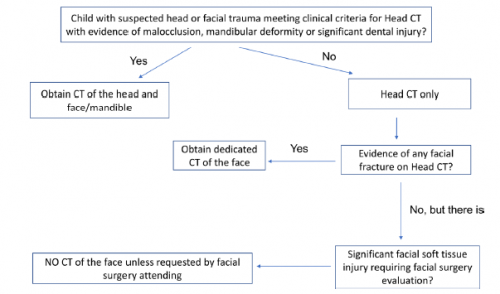
Here is the algorithm the authors recommend. Although designed for children, it should work for adults just as well.

Reference: Clinical and radiographic predictors of the need for facial CT in pediatric blunt trauma: a multi-institutional study. Trauma Surg Acute Care Open 2022;7:e000899.

Best Of EAST #8: Timing Of Reimaging For BCVI

There are still many questions regarding optimal management of blunt carotid and vertebral arterial injury (BCVI). We know that they may ultimately result in a stroke. And we kind of know how to manage them to try to avoid this. We also know that the grade may change over time, and many vascular surgeons recommend re-imaging at some point.

But when? There are still many questions. A multi-center trial has been collecting observational data on this issue since 2018. The group reviewed three years of data to examine imaging characteristics and stroke rate during the study period.

Here are the factoids:

  • A total of 739 cases were identified at 16 trauma centers
  • The median number of imaging studies was 2, with a range of 1-9 (!). Two thirds received only one study.
  • Injury grade distribution was as follows:
    • Grade 1 – 42%
    • Grade 2 – 30%
    • Grade 3 – 10%
    • Grade 4 – 18%
    • Grade 5 – <1%
  • About 30% changed in grade during the hospitalization, with 7% increasing and 24% decreasing.
  • Average time to change in grade was 7 days
  • Nearly 75% of those that decreased actually resolved. All of the grade 1 lesions resolved.
  • Stroke tended to occur after about one day after admission, although the grade 1 lesions took longer at 4 days
  • Strokes occurred much earlier than grade change

The authors concluded that there should be further investigation about the utility of serial imaging for stroke prevention.

Bottom line: This is basically a “how we did it” study to tease out data on imaging and stroke after BCVI. It’s clear that there is no consensus across trauma centers regarding if and when repeat imaging is done. And it’s not really possible to make any recommendations about repeat imaging based on this study.

However, it does uncover one important fact. It takes a week for the injury grade on CT to change, but strokes occur much earlier and usually within 24 hours! This is important because it makes it clear that it’s crucial to actually make the diagnosis early. Average stroke occurrence was 9% overall. Grade 1 injuries had only a 3% rate, but grades 2-4 were in the 12-15% range. Grade 5 had a 50% stroke rate!

These facts reinforce the importance of identifying as many of these BCVI as possible during the initial evaluation. The abstract I reviewed yesterday confirmed that the existing screening criteria (Memphis, Denver) will miss too many. More liberal imaging is probably indicated. If you missed the post, click here to view it in a new window.

Here are my comments for the authors and presenter:

  • The “change in BCVI grade over time” charts in the abstract are not readable. Please provide clear images during your presentation and explain what they mean. I was confused!
  • Based on your data, do you have any recommendations regarding the utility of re-imaging? Is it necessary in the same hospitalization at all? These patients will receive treatment anyway, and it doesn’t appear to have any impact on stroke rate.
  • Do you have any recommendations regarding the (f)utility of existing screening systems given the early occurrences of stroke in the study? Are you a fan of using energy / mechanism rather than a bullet list of criteria?

This is important work and I can’t wait to look at the data up close.

Reference: BLUNT CEREBROVASCULAR INJURIES: TIMING OF CHANGES TO INJURY GRADE AND STROKE FORMATION ON SERIAL IMAGING FROM AN EAST MULTI-INSTITUTIONAL TRIAL. EAST 35th ASA, oral abstract #34.