Category Archives: Anatomy

What Is: The Tripod Fracture?

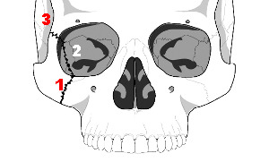
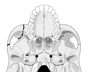
The tripod fracture (officially known as the zygomaticomaxillary complex fracture, and sometimes called a malar fracture) is the most common one seen after trauma. Fundamentally, the zygoma is separated from the rest of the face in a tripod fracture.

As you might imagine (tripod fracture), there are three components to this fracture. The first is a fracture through the zygomatic arch (1). Next, the fracture extends across the floor of the orbit and includes the maxillary sinus (2). Finally, the fracture includes the lateral orbital rim and wall (3).

Extraocular muscles may become trapped in the fracture line, leading to diplopia. It is very important to do a good eye exam to try to detect entrapment. The infraorbital nerve also passes through the orbital floor and may be injured, leading to numbness along the lower eyelid and upper lip.

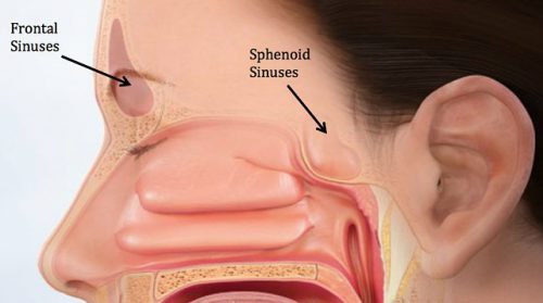
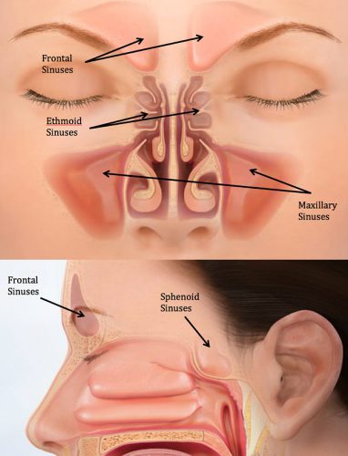
Nondisplaced fractures are treated symptomatically and reevaluated after a week or so to see if surgery would be beneficial. Displaced or symptomatic fractures require early open reduction. The pictures below show the anatomy of these fractures. They are derived from teaching materials provided by the Radiology Department at the University of Washington.

image

image

Print Friendly, PDF & Email

Zebra Alert: Blunt Injury To The Thoracic Duct

Today I’m going to review a very uncommon clinical problem in trauma: injury to the thoracic duct. To review, the lymphatic system coalesces into channels along the spine. These vessels travel upwards to drain into the venous system as the left and right lymphatic ducts. The exact drainage points vary, but are near the junctions of the internal jugular and subclavian veins bilaterally. Technically the larger of the two, the left lymphatic duct, is termed the thoracic duct. However, injuries can occur on either side.

Injuries to the lymphatic ducts are very rare due to their small size and their protection by surrounding structures and tissues. For this reason, the literature on this topic consists almost exclusively of case reports. Injury can occur from direct penetration by gunshots or stabs, or may be associated with high energy blunt trauma. It has also been reported to occur in cases of multiple posterior rib fractures and vertebral fractures.

In the rare event that these ducts are damaged, they pose a major management problem because lymph does not clot. These vessels are not self-sealing like most others in the body. They will only close through healing (scarring) or by ligation. The typical disruption occurs near the junction of the duct with the venous system, so lymph (chyle) typically accumulates in the thorax on the affected side. This results in a hydrothorax until the patient begins eating, when it turns chylous and makes the diagnosis easy. Here a various shades of chyle that you might see in the chest tube drainage.

If in doubt, triglyceride levels can be measured, and a value greater than 110 mg/dL is considered positive.

Initial management is usually dietary, via reduction in fat intake to render the drainage clear. This may be accomplished by a low fat diet or by TPN. I don’t really buy the effectiveness of this, since the fat content is not what causes the leak to persist. It merely makes it unusual to look at. I suspect that the 1-2 week period that most recommend for dietary treatment just provides an opportunity for normal healing/scarring to occur. Octreotide should be given as well because it may decrease overall lymphatic output. Lower output accelerates closure because the amount of scarring needed to close the smaller hole is less.

Interventional radiologists have attempted embolization and needle maceration of the ducts, but the few of these described have been unsuccessful. This is not recommended.

If closure is not achieved in two weeks, then consideration should be given to surgical ligation of the leaking duct. This structure is small and thin-walled, and not the easiest to see. Fats should be administered via NG (olive oil and cream have been described) at the start of the operation to stimulate chyle production. This allows easier identification of the leak site intraoperatively. Suture ligation, clipping, or both can be used to stop the leak.

References:

  1. A case of a traumatic chyle leak following an acute thoracic spine injury: successful resolution with strict dietary manipulation. World J Emerg Surg 6:10, 2011.
  2. Blunt rupture of the thoracic duct after severe thoracic trauma. J Trauma Open 3:e000183. doi:10.1136/tsaco-2018-000183m 2018.
  3. Bilateral Chylhotorax after Falling from Height. Case Reports in Surg  article 618708, 2014.
Print Friendly, PDF & Email

What You Need To Know About Frontal Sinus Fractures

Fracture of the frontal sinus is less common than other facial injuries, but can be more complex to deal with, both in the shorter and longer terms. These are generally high energy injuries, and facial impact in car crashes is the most common mechanism. Fists generally can’t cause the injury, but blunt objects like baseball bats can.

Here’s the normal anatomy:

sinus-fracture-treatment


Source: www.facialtraumamd.com

There are two “tables”, the anterior and the posterior. The anterior is covered with skin and a small amount of subcutaneous tissue. The posterior table is separated from the brain by the meninges.

Here’s an image of an open fracture involving both tables. Note the underlying pneumocephalus.

frontal_sinus1

A third of injuries violate the anterior table, and two thirds violate both. Posterior table fractures are very rare. A third of all patients will develop a CSF leak, typically from their nose.

These fractures may be (rarely) identified on physical exam if deformity and flattening is noted over the forehead. Most of the time, these patients undergo imaging for brain injury and the fracture is found incidentally. Once identified, go back and specifically look for a CSF leak. Clear fluid in the nose is, by definition, CSF. Don’t waste time on a beta-2 transferring (see below).

If a laceration is clearly visible over the fracture, or if a CSF leak was identified, notify your maxillofacial specialist immediately. If more than a little pneumocephalus is present, let your neurosurgeon know. Otherwise, your consults can wait until the next morning.

In general, these patients frequently require surgery for the fracture, either to restore cosmetic contours or to avoid mucocele formation. However, these are seldom needed urgently unless the fracture is an open fracture with contamination or there is a significant CSF leak. If in doubt, though, consult your specialist.

Related posts:

Print Friendly, PDF & Email

Sinus Precautions: Another Trauma Urban Legend?

The trauma service typically helps our facial surgery colleagues manage patients from time to time. Every now and again, I see their request/instructions for “sinus precautions” when patients have certain fracture patterns or undergo surgery involving sinuses.  Where did these “precautions” come from? Do they really make sense?

The paranasal sinuses shown above are the most commonly injured after facial trauma. They are lined with mucosa, and like the rest of our body, colonized with bacteria. They are interconnected with each other and the nasal cavity via tiny ostia that allow for normal drainage into the nose. Facial surgeons worry that these bacteria may seep out of fractured areas into clean tissue and cause deep infections. This may occur spontaneously, and could be accelerated if the pressure in the sinuses is increased in any way.

If you do a simple internet search on “sinus precautions”, it seems like every facial surgery group in the country has a set in their patient information sheet. And they all look suspiciously similar. As if they’ve been copied from each other and over, and from the same very, very old document.

As noted above, the main issue to be avoided is increasing pressure across the sinuses. Here’s the usual list of precautions, and my comments on their utility.

  • Avoid blowing your nose. This one actually makes sense. Closing the nose and mouth, pressurizing the pharynx and releasing through the nose will certainly increase sinus pressures. And there are actually reported cases of new onset cellulitis after this maneuver. True
  • Avoid sneezing. Good luck on this one. Who actually does this on purpose anyway? Sneezing will certainly increase sinus pressures, but this can be minimized by avoiding trying to stifle the sneeze. True
  • Do not drink through a straw. What? This causes a low pressure zone in the oropharynx so that fluids can be sipped, but the nasopharynx is isolated. If it weren’t you’d have beverages pouring out of your nose. False
  • Do not smoke. Well, this is certainly a good idea for many reasons, but has little impact on nasopharyngeal pressures. If anything, it decreases them slightly on inhalation, and works the same on exhalation as breathing out without the smoke. False
  • Do not blow musical instruments, balloons, … Yes, this can increase pressures. True
  • Do not push or lift heavy objects.  Hmm. The natural tendency is to perform a Valsalva maneuver (holding breath against a closed glottis) when doing this. In theory, this shouldn’t impact the sinuses because they are not attached to the trachea in any way. And even though it is possible to let some air escape the glottis and keep your mouth closed, it immediately exits the nose. Exhaling through the nose rapidly could increase pressures slightly. Most people don’t do this. Mostly false
  • Do not bend over, keep your head above your heart, sleep with your head slightly elevated. Come on, now. Sinuses are rigid, air-filled cavities. They don’t compress. False
  • Do not fly in a plane.  Definitely an issue. Everybody experiences popping ears and sinuses when flying. And it’s the descent that is of most concern. The increasing air pressure during this phase of flight can push fluid and air out of the sinuses. True
  • Do not spit. Okay, this goes without saying. It’s rude, but doesn’t do a thing to your sinuses. False

Bottom line: I was unable to find any seminal paper or book chapter as the source for “sinus precautions”. Most of the items on the list are bogus. But a few do actually increase sinus pressures and could result in fluid, air, and bacteria moving out of sinuses and into areas where they don’t belong. Pare down the list before you hand it to your patients.

Print Friendly, PDF & Email

Trauma Residents: How To Remember Liver Anatomy

In trauma surgery, operative management of liver injury is usually messy business, with little time for nice anatomic resections. However, an understanding of the basic anatomy, especially that of the vascular supply is crucial for saving your patient.

A cool tool for remembering Couinaud’s segments and the overall layout of liver anatomy was published in the Archives of Surgery recently. It makes use of a model, which consists of your hand! Just make a fist with your right hand and tuck the thumb behind the other fingers.


The fingers can then be numbered according to the Couinaud segments, with the caudate lobe (segment 1) represented by the thumb that is tucked away. The PIP joints represent the plane that the portal vein runs through, with branches going to upper and lower segments. Note how the ring finger normally lies a little more anterior than the little finger in this position, just like the sectors of the right lobe.

The creases between the fingers represent the left, middle and right hepatic veins.


The right hepatic vein is located between the right anterior and posterior sectors and the left hepatic vein sits between the left medial and lateral sectors. The middle hepatic vein is in between the left and right hemi-liver.

Bottom line: This “handy” liver model is available immediately in the OR and is already sterile. It can help visualize liver structures that may be injured quickly and accurately to speed your operative approach to the problem.

Reference: A Handy Tool to Teach Segmental Liver Anatomy to Surgical Trainees. Arch Surg 147(8):692-693, 2012.

Print Friendly, PDF & Email