Category Archives: Anatomy

EAST 2017 #10: A Simple Way To Predict Complications After Rib Fracture?

Rib fractures are a common injury, and a very common cause of morbidity. Every time I admit an elderly patient with rib fractures, I debate whether they should go to the ICU or a ward bed. Could there be a more objective way of determining the likelihood of complications, aggressiveness of treatment, and admission unit?

A group at West Virginia University implemented a rib fracture pathway in 2009, and have been collecting data on patients ever since. It was based on the measurement of forced vital capacity (FVC) on admission. This is the total amount of air that can be exhaled during a forced breath.

The authors subdivided their patients into two groups based on the total volume exhaled (<1.5L, and >1.5L). They retrospectively reviewed 6 years of data, looking at specific injuries, complications, and unexpected transfer to ICU. They hypothesized that patients in the highest FVC group would have fewer complications.

Here are the factoids:

  • There was a nearly even split in groups, with 678 patients who had FVC > 1.5L, and 682 with FVC < 1.5
  • There were significantly fewer complications and pneumonia, as well as fewer readmissions in the FVC > 1.5 group
  • Higher FVC was not associated with fewer unexpected transfers to ICU
  • Length of stay was half as long (4d vs 8d) in the high FVC group, but no p value was provided
  • The authors conclude that patients with FVC much greater than 1.5 are at lower risk for complications regardless of the number of fractures (???!)
  • They even suggest that patients with FVC > 1.5 could be discharged from the ED rather than be admitted (!)

Bottom line: Well, it started out good! The abstract showed that the high FVC patients had fewer complications and readmissions. And the length of stay was shorter, although significance was not noted. But the jump to correlating complication risk with number of fractures was not addressed in the abstract. And I can’t quite grasp the leap to suggesting possible discharge from the ED. 

FVC may be an inexpensive and simple test to administer in new rib fracture patients. But it’s ability to predict who goes to ICU and who goes home from the ED was not really identified in the study. 

Questions and comments for the authors/presenters:

  1. A minor point, but the upper limit was defined as > 1.5L in some parts of the abstract, and > 1.5L in  others. Small point, but keep it clean. Make sure all the greater than, less than, and equals signs are consistent.
  2. Was the shorter length of stay significantly different between the groups?
  3. Did you do any stratification by age?
  4. How did you make the conclusion that patients could be sent home from the ED?
  5. And did you do any correlations with your FVC data and the number of fractures? It’s not in the abstract.

Click here to go the the EAST 2017 page to see comments on other abstracts.

Related post:

Reference: Is an FVC of 1.5 adequate for predicting respiratory sufficiency in rib fractures? Paper #4, EAST 2017.

Print Friendly, PDF & Email

Why Do They Call It: The Surgical Neck of the Humerus?

Anatomy is complex and confusing at times. Pretty much everything you can find in the human body has a name. Sometimes it makes sense. Sometimes it’s named after someone famous. And sometimes, it’s just a head-scratcher.

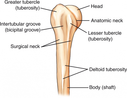
Let’s take the surgical neck of the humerus. Here’s an image of the proximal humerus:

proximal_humerus-14a181ca9b3646a88cc1

Notice there are two different “necks” of the humerus. You are probably familiar with the anatomic neck from your anatomy classes. But if you are a resident, an orthopedic surgeon, or someone who deals with fractures regularly, you are more familiar with the surgical neck.

The surgical neck of the humerus is the most common fracture site on the proximal humerus.  But here’s the kicker. It’s a misnomer!

Just because you see a fracture of the surgical neck of the humerus doesn’t meed it needs surgery! Indeed, many of these fractures are now successfully treated with immobilization in a sling. Your friendly neighborhood orthopedic surgeons will assess fracture stability by looking at the mechanism, exact location, involvement of the tubercles, and motion. Then they will decide on their treatment plan.

Bottom line: Don’t get suckered when someone asks you what operation is usually needed for a fracture of the surgical neck of the humerus!

Related posts: 

Print Friendly, PDF & Email

What You Need To Know About Frontal Sinus Fractures

Fracture of the frontal sinus is less common than other facial injuries, but can be more complex to deal with, both in the shorter and longer terms. These are generally high energy injuries, and facial impact in car crashes is the most common mechanism. Fists generally can’t cause the injury, but blunt objects like baseball bats can.

Here’s the normal anatomy:

sinus-fracture-treatment


Source: www.facialtraumamd.com

There are two “tables”, the anterior and the posterior. The anterior is covered with skin and a small amount of subcutaneous tissue. The posterior table is separated from the brain by the meninges.

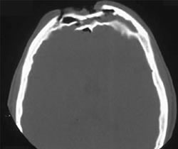
Here’s an image of an open fracture involving both tables. Note the underlying pneumocephalus.

frontal_sinus1

A third of injuries violate the anterior table, and two thirds violate both. Posterior table fractures are very rare. A third of all patients will develop a CSF leak, typically from their nose.

These fractures may be (rarely) identified on physical exam if deformity and flattening is noted over the forehead. Most of the time, these patients undergo imaging for brain injury and the fracture is found incidentally. Once identified, go back and specifically look for a CSF leak. Clear fluid in the nose is, by definition, CSF. Don’t waste time on a beta-2 transferring (see below).

If a laceration is clearly visible over the fracture, or if a CSF leak was identified, notify your maxillofacial specialist immediately. If more than a little pneumocephalus is present, let your neurosurgeon know. Otherwise, your consults can wait until the next morning.

In general, these patients frequently require surgery for the fracture, either to restore cosmetic contours or to avoid mucocele formation. However, these are seldom needed urgently unless the fracture is an open fracture with contamination or there is a significant CSF leak. If in doubt, though, consult your specialist.

Related posts:

Print Friendly, PDF & Email

The Chance Fracture

Centers that take care of blunt trauma are familiar with the spectrum of injury that is directly attributable to seat belt use. Although proper restraint significantly decreases mortality and serious head injury, seat belts can cause visceral injury, especially to small bowel.

Lap belt use has been associated with Chance fracture (flexion distraction injury to the lumbar spine) since 1982. The association between seat belts and intra-abdominal injury, especially with an obvious “seat belt sign” was first described in 1987.

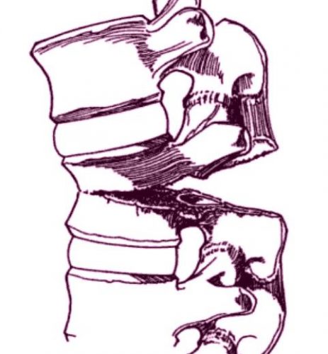
chance-fracture-21

Chance fracture. The vertebra appears to split in half from posterior to anterior.

Twenty years ago, orthopedic surgeons in Manitoba finally put two and two together and reported a series of 7 cases of Chance fractures. They noted that 6 of the fractures were associated with restraint use. Seat belt sign was also present in 5 of the 6 patients with fractures and three of the six had bowel injuries.

The authors noted that many provinces were mandating seatbelt use at the time, and they predicted that the number of Chance fractures, seat belt signs and hollow viscus injuries would increase. On the positive side, the number of deaths and serious head injuries would be expected to decline.

Although this was a small series, it finally cemented the unusual Chance fracture, seat belt sign, and bowel injury after motor vehicle trauma.

Thankfully, three point restraints (lap belt + shoulder harness) has been required in the seats next to doors for a long time. And since 2007, they have been mandated in the middle seat as well. Thus, these injuries seldom occur in any but the oldest (beater) cars on the road. They are seen more frequently now with sports and extreme sports injuries.

Chance fractures are frequently unstable, involving all three columns of the spine. The anterior column fails under compression, and the middle and posterior columns fail from the distraction mechanism. Usually, this fracture pattern requires operative fixation. However, if the posterior column is intact, a TLSO brace can be tried. This fracture is at risk for non-union and development of kyphosis or a flat back, which can lead to chronic pain and an abnormal posture.

Reference: Pediatric Chance Fractures: Association with Intra-abdominal Injuries and Seatbelt Use. Reid et al. J Trauma 30(4) 384-91, 1990.


Print Friendly, PDF & Email

The Cardiac Box: Meaningful For Gunshot Wounds?

A common dogma in trauma training is: “Watch out for the box!” This area on the anterior chest is purported to indicate high risk of cardiac injury in patients with penetrating trauma.

Where is it, exactly? Technically, it’s the zone extending from nipple to nipple, and from sternal notch to xiphoid.

The cardiac box

But is the dogma true? A number of (old) papers mapped out the location and incidence of cardiac injury in stabs to the chest and upper abdomen. And there is a pretty good correlation. For stab wounds. But what about gunshots?

A team at Emory University ran a retrospective review of their trauma registry data over a three year period.

Here are the factoids:

  • They saw nearly 90 patients per year with penetrating chest wounds. Of these, 80% were gunshots (!) Many had more than one penetration.
  •  Of the 233 gunshots inside “the box”, 34% injured the heart
  • The remaining 44 gunshots outside “the box” hit the heart 32% of the time
  • The authors suggest shifting the definition of “the box” toward the left, so that it extends from anterior midline, wraps around the left chest, and ends in the posterior midline (see below)

new-cardiac-box

Bottom line: Here’s the problem. Knives are attached to a handle which tends to stay outside your patient. Thus, it can only go so deep. But a bullet will keep going until something stops it, or it runs out of gas. So it makes sense that the traditional boundaries of “the box” don’t apply. But extending it to include the left lateral chest and exclude everything on the right side? It may make statistical sense in this study, but common sense dictates that the trauma professional needs to think about the heart any time a gunshot goes anywhere near the chest or upper abdomen. Do not limit yourself to any “box!”

Reference: Redefining the cardiac box: evaluation of the relationship between thoracic gunshot wounds and cardiac injury. AAST 2016 Paper #12.

Print Friendly, PDF & Email