Category Archives: Anatomy

Who Is At Risk For Blunt Cerebrovascular Injury?

Yesterday, I wrote about proper screening for blunt cerebrovascular injury (BCVI). But, as you know, it’s important to screen only when there is a significant risk of the injury being present. Screening using the shotgun approach (screen everyone for everything) yields enough false positive results to present potential danger to your patient.

A variety of authors on this topic have promoted a number of high risk criteria to trigger a screening test. Most make sense, and are related to the anatomy of the vessels in question. The carotid arteries are relatively unprotected, although a bit deep, as they course up the neck. Thus, it is possible to damage them when they suffer a direct and significantly hard blow. Once they enter the skull, they are better protected. However, fractures through key areas of the skull base and face can injure the vessels, even in these protected locations.

The vertebral arteries are deep and relatively protected as they course through the vertebral foramina. However, if the vertebrae are fractured or subluxed, vessel injury can occur.

Finally, and as always, the physical exam is important. If there are unexpected neurologic changes that can’t be explained by other injuries, or there are indications of deep vascular injury, BCVI needs to be considered.

Here is my list of indications to screen for BCVI:

  • Neurologic abnormality not explained by diagnosed injury
  • Arterial epistaxis†
  • Seat belt sign on neck†
  • GCS < 8 (this is the most commonly forgotten one)
  • Petrous bone fracture
  • C‐spine fracture (C1‐C3) or subluxation at any level†
  • Fracture through foramen transversum†
  • LeFort II or III fractures

Bottom line: Be on the lookout for any of the criteria listed above in your trauma patient. If you find one during your initial evaluation, be sure to order a CT angiogram of the neck. And keep an eye out while scanning the head and cervical spine. If any of the other radiographic indications become apparent, add on the CT angiogram at that point.

Print Friendly, PDF & Email

Bucket Handle Injuries Of The Intestine

Bucket Handle Injury

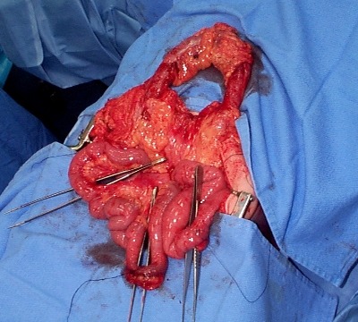
A bucket handle injury is a type of mesenteric injury of the intestine. The intestine itself separates from the mesentery, leaving a devascularized segment of bowel that looks like the handle on a bucket (get it?).

These injuries can occur after blunt trauma to the abdomen. The force required is rather extreme, so the usual mechanism is motor vehicle crash. In theory, it could occur after a fall from a significant height, and I have seen once case where a wood fragment was hurled at the abdomen by a malfunctioning lathe.

The mechanics of this injury are related to fixed vs mobile structures in the abdomen. Injuries tend to occur adjacent to areas of the intestine that are fixed, such as the cecum, ligament of Treitz, colonic flexures and rectum. During sudden deceleration, portions of the intestine adjacent to these areas continue to move, pulling on the nearby attachments. This causes the intestine itself to pull off of its mesentery.

The terminal ileum is the most common site for bucket handle tears. Proximal jejunum, transverse colon, and sigmoid colon are other possible areas. The picture above shows multiple bucket handle injuries in one patient. There are 3 injuries in the small bowel, and one involving the entire transverse colon. Note the obviously devascularized segment at the bottom center of the photo.

Always think about the possibility of this injury in patients with very high speed decelerationsSeat belt marks have a particularly high association with this injury. If your patient has an abnormal exam in the right lower quadrant, or if the CT shows unusual changes there (“dirty” mesenteric fat, thickened bowel wall, extravasation), I recommend a trip to the OR. In these cases, an injury will nearly always be present.

Related posts:

Source: personal archive. Not treated at Regions Hospital

Print Friendly, PDF & Email

Trauma Residents: How To Remember Liver Anatomy

In trauma surgery, operative management of liver injury is usually messy business, with little time for nice anatomic resections. However, an understanding of the basic anatomy, especially that of the vascular supply is crucial for saving your patient.

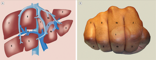
A cool tool for remembering Couinaud’s segments and the overall layout of liver anatomy was published in the Archives of Surgery recently. It makes use of a model, which consists of your hand! Just make a fist with your right hand and tuck the thumb behind the other fingers.


The fingers can then be numbered according to the Couinaud segments, with the caudate lobe (segment 1) represented by the thumb that is tucked away. The PIP joints represent the plane that the portal vein runs through, with branches going to upper and lower segments. Note how the ring finger normally lies a little more anterior than the little finger in this position, just like the sectors of the right lobe.

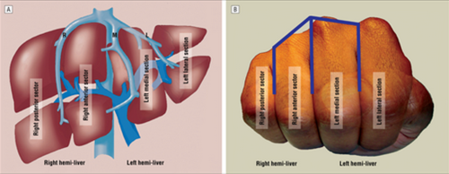
The creases between the fingers represent the left, middle and right hepatic veins.


The right hepatic vein is located between the right anterior and posterior sectors and the left hepatic vein sits between the left medial and lateral sectors. The middle hepatic vein is in between the left and right hemi-liver.

Bottom line: This “handy” liver model is available immediately in the OR and is already sterile. It can help visualize liver structures that may be injured quickly and accurately to speed your operative approach to the problem.

Reference: A Handy Tool to Teach Segmental Liver Anatomy to Surgical Trainees. Arch Surg 147(8):692-693, 2012.

Print Friendly, PDF & Email

Bucket Handle Injury – Part 2

Yesterday, I wrote about the basics of bucket handle injuries of the intestine. Today, I’ll deal with diagnosing them.

An understanding of the mechanism of injury and a good physical exam are paramount. If the patient took a significant blow to the abdomen, especially in a motor vehicle crash (lap belt), be very suspicious. Any abdominal pain is of concern, particularly in the right lower quadrant (most common injury location). If a CT is indicated and there are focal changes in the mesenery or bowel wall, a trip to the OR is advised.

In some patients, the bowel is devascularized and takes 2-3 days to become necrotic. They experience slowly increasing focal pain, and once this develops it’s time to go to the operating room.

Intubated and/or comatose patients can be problematic in making this diagnosis. There is no physical exam, so the trauma professional has to rely on surrogates. The white blood cell (WBC) count is very helpful. The WBC count is typically elevated into the 15,000-20,000 range immediately after trauma, and declines to normal within about 12 hours. If it begins to climb again after 24 hours, especially if it exceeds 20,000, an intestinal injury is likely.

CT scan and abdominal ultrasound are also helpful. A repeat CT scan may show a change in the volume of fluid, or a change in its character. If the amount of fluid increases significantly, or if a fluid bi-layer is seen, a bucket handle injury is very likely. These findings are pertinent in awake patients as well, but the physical exam usually makes use of these diagnostics unnecessary.

Related posts:

Print Friendly, PDF & Email

Bucket Handle Injury – Part 1

Bucket Handle Injury

A bucket handle injury is a type of mesenteric injury of the intestine. The intestine itself separates from the mesentery, leaving a devascularized segment of bowel that looks like the handle on a bucket (get it?).

These injuries can occur after blunt trauma to the abdomen. The force required is rather extreme, so the usual mechanism is motor vehicle crash. In theory, it could occur after a fall from a significant height, and I have seen once case where a wood fragment was hurled at the abdomen by a malfunctioning lathe.

The mechanics of this injury are related to fixed vs mobile structures in the abdomen. Injuries tend to occur adjacent to areas of the intestine that are fixed, such as the cecum, ligament of Treitz, colonic flexures and rectum. During sudden deceleration, portions of the intestine adjacent to these areas continue to move, pulling on the nearby attachments. This causes the intestine itself to pull off of its mesentery.

The terminal ileum is the most common site for bucket handle tears. Proximal jejunum, transverse colon, and sigmoid colon are other possible areas. The picture above shows multiple bucket handle injuries in one patient. There are 3 injuries in the small bowel, and one involving the entire transverse colon. Note the obviously devascularized segment at the bottom center of the photo.

Always think about the possibility of this injury in patients with very high speed decelerations. Seat belt marks have a particularly high association with this injury. If your patient has an abnormal exam in the right lower quadrant, or if the CT shows unusual changes there (“dirty” mesenteric fat, thickened bowel wall, extravasation), I recommend a trip to the OR. In these cases, an injury will nearly always be present.

Tomorrow: These injuries can be subtle in an awake patient with a reliable exam. On Friday I’ll write about how you can detect it in unconscious patients.

Source: personal archive. Not treated at Regions Hospital

Print Friendly, PDF & Email