Category Archives: Mechanism

Closing Velocity And Injury Severity

Trauma professionals, both prehospital and in trauma centers, make a big deal about “closing velocity” when describing motor vehicle crashes.  How important is this?

So let me give you a little quiz to illustrate the concept:

Two cars, of the same make and model, are both traveling on a two lane highway at 60 mph in opposite directions. Car A crosses the midline and strikes Car B head-on. This is the same as:

  1. Car A striking a wall at 120 mph
  2. Car B striking a wall at 60 mph
  3. Car A striking a wall at 30 mph

2010-saab-9-5-head-on-crash-test_100313384_m1

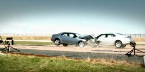
The closing velocity is calculated by adding the head-on components of both vehicles. Since the cars struck each other exactly head-on, this would be 60+60 = 120 mph. If the impact is angled there is a little trigonometry involved, which I will avoid in this example. And if there is a large difference in mass between the vehicles, there are some other calculation nuances as well.

So a closing velocity of 120 mph means that the injuries are worse than what you would expect from a car traveling at 60 mph, right?

Wrong!

In this example, since the masses are the same, each vehicle would come to a stop on impact because the masses are equal. This is equivalent to each vehicle striking a solid wall and decelerating from 60 mph to zero immediately. Hence, answer #2 is correct. If you remember your physics, momentum must be conserved, so both of these cars can’t have struck each other at the equivalent of 120 mph. The injuries sustained by any passengers will be those expected in a 60 mph crash.

If you change the scenario a little so that a car and a freight train are traveling toward each other at 60 mph each, the closing velocity is still 120 mph. However, due the the fact that the car’s mass is negligible compared to the train, it will strike the train, decelerate to 0, then accelerate to -60 mph in mere moments. The train will not slow down a bit. For occupants of the car, this would be equivalent to striking an immovable wall at 120 mph. The injuries will probably be immediately fatal for all.

Bottom line: Closing velocity has little relationship to the injuries sustained for most passenger vehicle crashes. The sum of the decelerations of the two vehicles will always equal the closing velocity. Those injuries will be consistent with the change in speed of the vehicle the occupants were riding, and not the sum of the velocities of the vehicles. 

Print Friendly, PDF & Email

Button Batteries: Part 2 – Getting Them Out

In my last post, I detailed how to suspect and image a button battery ingestion. In this one, I’ll describe how to extract them, and how quickly it’s necessary.

When batteries come to rest and are surrounded by moist mucosal tissue, a current arc is generated around the two sides of the button. This releases heat, which coagulates the surrounding tissue. Depending on the location, closeness of contact, and the duration, these burn injuries may extend into underlying tissue. This is of particular significance in the esophagus, which is in close proximity to the thoracic aorta.

Here’s a simple demonstration you can do at home with some lunch meat:

Here are guidelines for what to do when you encounter pediatric patients who have ingested a button battery:

  • If the child is experiencing bleeding from the upper GI tract, activate your trauma team. The child may have an aorto-esophageal fistula. If there is no active bleeding, obtain a chest x-ray to assess the battery’s position. If there is active bleeding, proceed to the OR (preferably a hybrid room if you have one) and use fluoro to locate the battery. If bleeding persists, call appropriate pediatric surgical specialists (surgery, CV surgery, GI), activate your massive transfusion protocol, and consider tamponade with a Blakemore tube (remember those?) or a urinary catheter if you don’t have one.
  • No bleeding from the upper GI tract? If the battery is large (>20mm) and/or the child is small (<5 years), and is lodged in the esophagus, proceed immediately to OR and remove endoscopically.
  • Batteries in the stomach are of less concern. They will generally pass if <20mm. A repeat x-ray after 48 hours should be obtained for larger batteries. If still in the stomach, they should be removed endoscopically. Smaller batteries will usually pass, and should be re-imaged after two weeks to confirm this.

References:

  • Button battery and magnet ingestions in the pediatric patient.  Curr Opin Pediatrics 30:653-659, 2018.
  • Management of ingested foreign bodies in children: a clinical report of the NASPGHAN Endoscopy Committee. J Pediatric Gastroenterol Nutr 60:562-574, 2015.
Print Friendly, PDF & Email

Button Batteries: Part 1

I know what you are saying. Button batteries? Trauma? Not too many adult trauma professionals have seen or heard of this. But those who care for pediatric patients should be very familiar. If the importance of this seemingly minor problem is ignored, the results can be catastrophic.

Kids eat stuff, and not just food. The smaller ones always seem to be putting things in their mouths. Foreign body ingestion (or insertion into other orifices) is a common presentation at pediatric emergency departments. Unfortunately, the fact that a battery has been eaten may not be appreciated by the parents.  The child may be brought in with  nonspecific GI or respiratory symptoms.

As soon as a battery ingestion is known or suspected, a two-view chest x-ray is needed. This should show both chest and upper abdomen in order to visualize both esophagus and stomach. Separate chest and abdominal images may be required if the child is too large for a single shot. Two views (AP and lateral) are important because the nature of the foreign body may not be appreciated if the battery is seen edge-on.

If you are fortunate enough to image the battery “face-on”, you may see a telltale halo sign. Because of the way these batteries are put together, there are two metal sides that have a slight difference in overlap.

You’ve made the diagnosis! So now what? And how quickly? I’ll deal with this in my next post.

Print Friendly, PDF & Email

Trauma Activation For Hanging: Yes or No?

In my last post, I discussed a little-reviewed topic, that of strangulation. I recommended activating your trauma team only for patients who met the physiologic criteria for it.

But now, what about hangings? There are basically two types. The judicial hanging is something most of you will never see. This is a precisely carried out technique for execution and involves falling a certain height while a professionally fashioned noose arrests the fall. This results in a fairly predictable set of cervical spine/cord, airway, and vascular injuries. Death is rapid.

Suicidal hangings are far different. They involve some type of ligature around the neck, but rarely and fall. This causes slow asphyxiation and death, sometimes. The literature dealing with near hangings is a potpourri of case reports, speculation, and very few actual studies. So once again, we are left with little guidance.

What type of workup should occur? Does the trauma team need to be called? A very busy Level I trauma center reviewed their registry for adult near-hangings over a 19 year period. Hanging was strictly defined as a ligature around the neck with only the body weight for suspension. A total of 125 patients were analyzed, and were grouped into patients presenting with a normal GCS (15), and those who were abnormal (<15).

Here are the factoids:

  • Two thirds of patients presented with normal GCS, and one third were impaired
  • Most occurred at home (64%), and jail hangings occurred in 6%
  • Only 13% actually fell some distance before the ligature tightened
  • If there was no fall, 32% had full weight on the ligature, 28% had no weight on it,  and 40% had partial weight
  • Patients with decreased GCS tended to have full weight on suspension (76%), were much more likely to be intubated prior to arrival (83% vs 0% for GCS 15), had loss of consciousness (77% vs 35%) and had dysphonia and/or dysphagia (30% vs 8%)
  • Other than a ligature mark, physical findings were rare, especially in the normal GCS group. Subq air was found in only 12% and stridor in 18%.
  • No patients had physical findings associated with vascular injury (thrill, bruit)
  • Injuries were only found in 4 patients: 1 cervical spine fracture, 2 vascular injuries, and 1 pneumothorax
  • 10 patients died and 8 suffered permanent disability, all in the low GCS group

Bottom line: It is obvious that patients with normal GCS after attempted hanging are very different from those who are impaired. The authors developed an algorithm based on the initial GCS, which I agree with. Here is what I recommend:

  • Do not activate the trauma team, even for low GCS. This mechanism seldom produces injuries that require any surgical specialist. This is an exception to the usual GCS criterion.
  • The emergency physician should direct the initial diagnosis and management. This includes airway, selection of imaging, and directing disposition. A good physical exam, including auscultation (remember that?) is essential.
  • Patients with normal GCS and minimal neck tenderness or other symptoms do not need imaging of any kind.
  • Patients with abnormal GCS should undergo CT scanning, consisting of a CT angiogram of the neck and brain with soft tissue images of the neck and cervical spine recons.
  • Based on final diagnoses, the patient can be admitted to an appropriate medical service or mental health. In the very rare case of a spine, airway, or vascular injury, the appropriate service can be consulted.

Reference: A case for less workup in near hanging. J Trauma 81(5):925-930, 2016.

Print Friendly, PDF & Email

Trauma Activation For Strangulation: Yes or No?

Trauma activation criteria generally fall into four broad categories: physiology, anatomy, mechanism of injury, and co-factors. Of these, the first two are the best predictors of patients who actually need to be assessed by the full trauma team. Many trauma centers include a number of mechanistic criteria, usually much to their chagrin. They typically end up with frequent team activations and the patient usually ends up have trivial injuries.

However, there are some mechanisms that just seem like they demand additional attention. Death of another occupant in the vehicle. Fall from a significant height. But what about a patient who has been strangled?

Unfortunately, the published literature gives us little guidance. This usually means that trauma centers will then just do what seems to “make sense.” And unfortunately, this frequently results in significant overtriage, with many patients going home from the emergency department.

Since there is little to know research to show us the way, I’d like to share my thoughts:

  • As a guiding principle, the trauma  team should be activated when the patient will derive significant benefit from it. And the benefit that the team really provides is speed. The team approach results in quicker diagnosis from physical exam and FAST. It gets patients to diagnostic imaging quicker, if appropriate. And gets them to the OR faster when it’s not appropriate to go to CT.
  • Activating for a strangulation mechanism alone is probably a waste of time.
  • Look at the patient’s physiology first. Are the vital signs normal? What is the GCS? If either are abnormal, activate.
  • Then check out the anatomy. If the patient has any voice changes, or has obvious discoloration from bruising, crepitus, or subcutaneous emphysema, call the team. They may suffer a deteriorating airway at any moment.

If physiologic and anatomic findings don’t trigger an activation, then standard evaluation is in order. Here are some things to think about:

  • A complete physical exam is mandatory. This not only includes the neck, but the rest of the body. Strangulation is a common injury from domestic violence, and other injuries are frequently present.
  • If there are any marks on the neck, CT evaluation is required. This includes soft tissue, CT angiography, and cervical spine evaluation. All three can be done with a single contrast-enhanced scan. The incidence of spine injury is extremely low with strangulation, but the spine images are part of the set anyway.
  • CT of the chest is never indicated. There is no possibility of aortic injury with this mechanism, and all the other stuff will show up on the chest x-ray, if significant enough for treatment.
  • Even if there are no abnormalities, your patient may need admission while social services arranges a safe place for their discharge. Don’t forget the social and forensic aspects of this injury. Law enforcement may need photographic evidence or statements from the patient so this event can’t happen again.

Next post: Trauma Activation for Hanging: Yes or No?

Reference: Strangulation forensic examination: best practice for health care providers. Adv Emerg Nurs J 35(4):314-327, 2013.

Print Friendly, PDF & Email