Category Archives: Thorax

Prehospital: What’s The Best Chest Seal For Sucking Chest Wounds?

The treatment of a “sucking chest wound” in the field has typically been with application of some type of occlusive dressing. Many times, a generic adhesive dressing is applied, typically the same kind used to cover IV sites. This is quick, easy, cheap, and readily available in the ambulance. But there is a danger that this could result in development of tension pneumothorax, because the dressing not only keeps air from getting in but also keeps any buildup of pneumothorax from getting out.

To avoid this, a number of vented products have been developed and approved by the US Food and Drug Administration (FDA). These devices have some sort of system to allow drainage of accumulating air or blood, typically a one-way valve or drainage channels. They also need to stick well to a chest wall, which may have blood or other fluids that might disrupt the seal completely.

The US Army has a strong interest in making sure the products they use for this purpose work exactly as promised. The US Army Institute of Surgical Research examined 5 currently FDA-approved products to determine their ability to adhere to bleeding chest wounds, and to drain accumulating air and/or blood from the pleural space. They developed an open chest wound with active bleeding in a swine model.

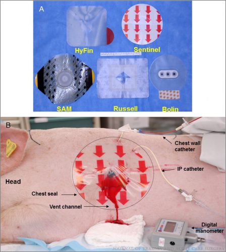
An open hemopneumothorax was created by infusing air and blood, the animal was stabilized, then additional aliquots of air and blood were infused to simulate ongoing bleeding and air buildup. The image below shows the 5 products used and the animal setup:

Here are the factoids:

  • Creation of the open hemopneumothorax caused the intrapleural pressure to move toward atmospheric pressure as expected, resulting in labored breathing and reduced O2 saturation
  • Sealing the wound with any of the chest seal products corrected all of the problems just noted
  • Chest seals with one way valves did not evacuate blood efficiently (Bolin and SAM). The dressings either detached due to pooled blood, or the vent system clogged from blood clot.
  • Seals with laminar channels for drainage (see the pig picture above) allowed easy escape of blood and air
  • Success rates were 100% for Sentinel and Russell, 67% for HyFin, 25% for SAM, and 0% for Bolin

Bottom line: Prehospital providers need to be familiar with the products they use to cover open chest wounds. Totally occlusive dressings can result in development of a tension pneumothorax if there is an ongoing air leak from the lung. Vented chest seals are preferable for these injuries. Just be aware that vented seals with drainage channels perform much better than those that rely on a one-way valve.

Reference: Do vented chest seals differ in efficacy? An experimental
evaluation using a swine hemopneumothorax model. J Trauma 83(1):182-189, 2017.

Print Friendly, PDF & Email

The “Backward Finochietto” Problem

Resuscitative thoracotomy is a (sometimes) life-saving procedure reserved for trauma patients in extremis. Thankfully, most trauma centers do very few of these a year. However, that makes it one of those “high severity – low frequency” procedures that generate many, many quality improvement problems. Many of these issues are due to operator unfamiliarity or equipment availability.

Today, I’ll highlight a problem that crops up occasionally at various trauma centers across the US: the “backward Finochietto.” One of the most essential components of the resuscitative thoracotomy is rapid access to the chest. A large skin incision is typically made, the thoracic wall and intercostal musculature are divided, and the pleural space is entered.

It’s not easy to insinuate your arm between the ribs in an average person. But, of course, there’s a retractor for that! Von Mikulicz presented the first rib spreader at a German surgical society meeting in 1904.  Various versions of this instrument were devised over the next three decades to make it easier and faster to use.

The Finochietto retractor was introduced in 1936 and boasted several enhancements. It used a rack and pinion system to make it easier for the surgeon to spread the chest wall and made it unlikely to close on its own. The turning lever was hinged so it could flattened and placed out of the surgical field. The blades contained fenestrations so chest wall tissue could protrude into them and keep it from slipping when opened. It remains a workhorse instrument for us today and is found in most instrument packs for resuscitative thoracotomy.

But there is a potential problem. Some Finochietto retractors consist of only two pieces: a blade with the linear gear teeth (the rack) and another blade that fits onto it with the turning handle (the pinion). See the image below:

Looks great, right? However, there is one downside. The retractor parts that hook into the soft tissue are of a fixed depth. What if your patient has a more generous body habitus? Placing multiple sets of this retractor into the thoracotomy pack is not practical.

The solution is to allow detachable blades of various sizes. Here’s a modern-day example:

The good news is that the retractor tips are interchangeable. The bad news is that they are sometimes interchangeable with the wrong arm of the retractor! Hence the “backward Finochietto” problem. It’s impossible to use the retractors with the blades on the wrong side, and it takes time the trauma professional does not have to figure out how to snap them off and switch them around.

So what’s the solution? This is clearly an instrument reprocessing quality issue. These instruments are expensive, so your hospital may not be excited about purchasing new ones just for the trauma bay. It all boils down to foolproofing it in as many ways as possible.  Here are some tips:

  • Provide an educational session for all of the reprocessing techs. Unfortunately, this effect will wear off as staff turnover occurs.
  • Post a photograph of a properly assembled retractor for the techs to use when processing the tray.
  • Use colored instrument marking tape on each piece of the instrument. For example, a green tape strip should be placed on both the rack arm of the retractor and the left blade. Use red tape for the pinion arm and the right blade. All the tech needs to do now is match the colors as they assemble the retractor.

Bottom line: This problem is more common than you may think. Ask one of your old-timer trauma surgeons and I’ll bet they can tell you some stories. But it is easily avoided with a little creativity and some tape! Be sure to do it now so it doesn’t pop up in the heat of a resuscitative thoracotomy .

Print Friendly, PDF & Email

What You Need To Know About Blunt Cardiac Injury

Blunt cardiac injury can be an enigma. Significant injuries are uncommon, and the literature on it consists of case reports and small series. The group at Scripps La Jolla has an excellent review article on the topic that is currently in press. This post will relate some of the key points in this nicely prepared article.

  • Use the correct nomenclature. For years, many have called this condition “cardiac contusion” or “myocardial contusion.” Unfortunately, these descriptors are too specific. The proper term is “blunt cardiac injury (BCI),” which encompasses the entire gamut of injury from asymptomatic contusion to pericardial injury to cardiac rupture.
  • BCI occurs more commonly than you think. If one defines BCI as any arrhythmia or cardiac enzyme elevation, it is fairly common. However, if the definition is limited to clinically significant sequelae such as potentially malignant arrhythmia or cardiac failure, the incidence is easily less than 1% in blunt trauma patients.
  • Be aware of the usual mechanisms of injury. This is a condition caused by blunt trauma, with motor vehicle crashes causing half and pedestrians struck by them another one-third. Motorcycle crashes and falls caused the remaining 12%.
  • Diagnosis can be challenging.
    • Physical examination is usually of little help. New onset of a heart murmur may indicate a serious cardiac injury but is exceedingly rare.
    • EKG evidence of a new onset arrhythmia is important, particularly bundle branch blocks, PVCs, and ST segment / T wave changes, which require further investigation.
    • CPK-MB enzyme measurements are useless. Please don’t get them.
    • Troponin T and Troponin I are frequently used but do not reliably predict BCI. Testing in asymptomatic patients is not helpful and may result in additional asymptomatic testing.
    • Echocardiography is not indicated in asymptomatic patients with isolated enzyme elevations.
    • Cardiac CT may be used to differentiate acute MI from BCI. Frequently, patients at risk are having a chest CT with contrast performed anyway.

Here is the recommended treatment algorithm:

  1. If BCI is possible based on mechanism of injury, follow the ATLS protocols and perform a physical exam, E-FAST, and place on EKG monitoring.
  2. If the patient is hemodynamically unstable, quickly identify and treat tamponade or tension pneumothorax if present. If significant arrhythmias are present, treat with appropriate medications. If heart failure is present, treat medically and evaluate for surgical problems such as valve, septum, or coronary artery injury.
  3. If the patient is hemodynamically stable, obtain a 12 lead EKG. If significant arrythmias are present, treat with appropriate medications. If there is organ hypoperfusion, obtain an echocardiogram. If this study reveals an effusion, a pericardial window is indicated. If the echo shows hypokinesis or structural injury, appropriate medical or surgical management should be carried out.
  4. Patients who have only significant arrhythmias should be admitted to a monitored bed for 24 hours. Once arrhythmias have resolved, the patent can be discharged.
  5. Patients with nonspecific EKG changes should have troponin levels drawn after 8 hours of observation in the ED. If elevated, admit to a monitored bed for 24 hours. Once EKG and troponin have normalized, the patent can be discharged.
  6. If EKG and labs are normal, may discharge home from the ED if there are no other indications for admission.

Reference: Diagnosis and Management of Blunt Cardiac Injury: What You Need to Know. J Trauma, accepted for publication. DOI: 10.1097/TA.0000000000004216

Print Friendly, PDF & Email

Early Vs Delayed Thoracic Endovascular Repair

Back in the day, the only way to fix a broken thoracic aorta was via left thoracotomy.  This was a big procedure, with the possibility of several major complications, with postop paraplegia being one of them. At the time, there was a debate about whether the procedure should be done immediately versus waiting until the patient was well-resuscitated. The concern was that death was nearly certain if the aortic lesion progressed.

We learned that temporizing with strict blood pressure control worked wonders at protecting the patient. Although many of these injuries were managed within hours, a growing number were delayed by a few days to improve outcomes.

Nowadays, thoracic endovascular aortic repair (TEVAR) is routine and much less morbid than the open procedure. However, the same question arises: do it early or wait a while? Interestingly, not one but two analyses have been published on this very topic in the last four months!

The first is from an international research group that searched the usual databases and initially found 921 records. They included only clinical trials or cohort studies with ten or more adult patients that could be stratified as early (within 24 hours) or late (after 24 hours) intervention. After applying these criteria, only seven studies remained for analysis.

There were 3,757 patients with early repairs, compared to 1,238 undergoing late repair. The presenting demographics and injury grades were similar in each group. However, the short-term mortality was significantly higher (1.9x) in the early TEVAR group. Additionally, ICU length of stay was significantly longer (3 days) in the late TEVAR group.

The second paper was presented as a quick-shot at last year’s AAST meeting. It is from a group of researchers from our big Boston trauma centers and the Netherlands. They used four years of data from the TQIP database, giving them extra information unavailable in the first study. They specifically looked at patients with grade II or III injuries. Here is the grading scale:

Here are the factoids:

  • A total of 1,339 patients were studied, with about three-quarters in the early TEVAR group
  • Median time to TEVAR was 4 hours in the early group and 65 hours in the late group
  • Patients in the early group were significantly less likely to have brain or liver injuries
  • ISS was similar in both groups
  • The early TEVAR group had significantly higher in-hospital mortality (16% vs. 5%), significantly higher risk of ARDS (7.6% vs. 2.1%), but significantly shorter ICU stay (7 vs 10 days)
  • When patients who died within the first 24 hours were excluded, the in-hospital mortality remained significantly higher, and the ICU and hospital lengths of stay were significantly shorter

Bottom line: Some society guidelines began recommending delayed TEVAR in 2015. This study did not detect any trend toward this, however. Using different methods and databases, these two studies identified nearly identical mortality and ICU trends in large groups of patients. The mortality trends do not appear to be related to injury grade, overall injury severity, or the presence of head injury. 

Taken together, this suggests that we need to rethink the timing of TEVAR in patients with grade II or III injuries. The best timing still needs to be defined, but it appears to be beyond 24 hours. Centers performing this procedure should review their results and consider extending procedure timing as additional research is done to define the ideal time interval.

References:

  1. Early Versus Delayed Thoracic Endovascular Aortic Repair for Blunt Traumatic Aortic Injury: A Systematic Review and Meta-Analysis. Cureus. 2023 Jun 28;15(6):e41078. doi: 10.7759/cureus.41078. PMID: 37519486; PMCID: PMC10375940.
  2. Early Versus Delayed Thoracic Endovascular Aortic Repair for
    Blunt Thoracic Aortic Injury: A Propensity Score-Matched Analysis. Ann Surg 278:e848-e854, 2023.


Print Friendly, PDF & Email

Best Of AAST #3: When To Place A Chest Tube For Hemothorax

There is an art to deciding when to place a  chest tube for either hemothorax or pneumothorax. For the most part, the trauma professional examines the imaging and then uses some unknown internal metric to declare that it is “too big.” Then it’s time to insert some type of chest drain.

There have been attempts over the years to make this decision more quantitative. One of the better-known ones is the 2-cm rule for pneumothorax. If the distance from the chest wall to the lung on the chest x-ray is >2cm, it is “too big.”

But what about hemothorax? The Medical College of Wisconsin trauma group performed a retrospective review of 391 patient charts to test a new 300cc rule defining when a hemothorax is “too big.” This guideline was implemented in 2018-2019, and patients presenting before implementation were compared to those arriving after.

The 300cc threshold is determined by using Mergo’s formula for calculating the volume of a square prism. Obviously, this requires a CT scan for calculation, so patients who had a tube placed before scanning or did not have one were excluded. They were also excluded from the study if their pneumothorax met the 2-cm rule. The authors studied how many patients could be observed, how many needed tube drainage, observation failure, and later need for a VATS procedure or thoracotomy.

Here are the factoids:

  • About 60% of the study group was admitted after the new criteria were implemented, and both groups were demographically similar
  • After implementation, the number of patients that were just observed increased significantly from 52% to 71%
  • Of course, this means that the number of chest tubes inserted was significantly less (42% vs. 61%)
  • There was no difference in observation failure (delayed placement of a tube), 18% vs. 24%
  • There were also no differences in pulmonary complications, 30-day readmissions, or 30-day mortality
  • The average ICU and hospital length of stays were significantly shorter as well

The authors concluded that implementing their 300cc guidelines correlated with decreased length of stay and no increase in failure or complication rates.

Bottom line: Although this is a relatively small series, the differences between the groups quickly achieved significance. There are three major questions that I have. First, how was the 300cc threshold arrived at? Was this borne of clinical judgment, or did some previous work suggest it?

My next question has to deal with the accuracy of the volume calculation. Mergo’s formula was used to determine the volume of a rectangular solid. As we all know, hemothoraces and pneumothoraces are not cubes. They can be very irregular and influenced by patient position. However, I did find a paper from the University of Florida that found the correlation coefficient between the volume calculated by Mergo’s formula vs. using 3-D software estimation was 0.9, which is excellent. So this approximation appears to be a very good one.

Finally, using the 300cc rule is predicated on getting a CT scan. Does every patient need a chest CT? Part of the resuscitation process for major trauma involves obtaining a chest X-ray. The obviously large hemothorax can justify inserting a chest tube at that point. But the reality is that most of these patients do go on to chest CT, so this is a minor change in practice for most. 

Although I love to see confirmatory studies before practice changes, this one study can lead us to change our practice guidelines now. It is a relatively minor one and will allow us to avoid placement of a few more chest tubes and to shave off a few days of hospital stay. The logical follow-up study for the authors is to extend the post-discharge window for complications to 60 or 90 days to ensure that delayed procedures were not required in the observation group.

References:

  • Implementing the 300-cc rule safely decreases chest tube placement in traumatic hemothorax. AAST 2023 Plenary paper #22.
  • New formula for quantification of pleural effusions from computed tomography. J Thorac Imaging. 1999 Apr;14(2):122-5. 


Print Friendly, PDF & Email