Category Archives: Thorax

Delayed Presentation Of Right Diaphragm Injury

Diaphragm injury from blunt trauma is uncommon, occurring in only a few percent of patients after high-energy mechanisms. They usually occur on the left side and are more frequently seen after t-bone type car crashes and in pedestrians struck by a car.

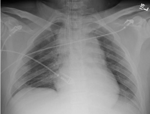
Blunt diaphragm injury on the right side is very unusual. Even so, it is more easily detected due to obvious displacement of the liver that can be seen on chest x-ray. Blunt injuries on the right side usually result in a large rent in the central tendon or detachment of the diaphragm from the chest wall. This allows the liver to herniate into the chest, and the chest x-ray finding is not subtle.

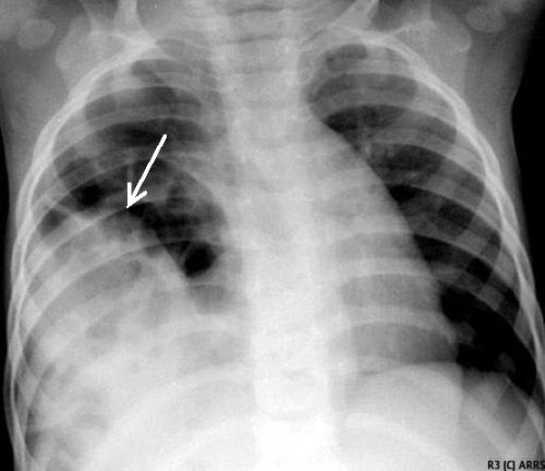
This image shows an acute herniation of the liver through the diaphragm. Due to the size of the liver, only part of it can typically fit through the rent. Radiologists call this the “cottage loaf” sign. Why? Here’s the bakery item it is named after. Get it now?

Thankfully, most of these injuries are identified in the acute setting. They must be addressed surgically because, if left untreated, more and more of the liver will slowly move into the chest resulting in respiratory problems in the long run.

Acute management usually consists of laparotomy to address both the diaphragm tear and any other associated intra-abdominal injuries. The liver should be reduced by sliding a hand next to it laterally into the chest cavity and pushing the dome downwards. The right triangular ligaments should be taken down (if they are not already destroyed) to mobilize the organ better so the diaphragm laceration can be closed. This is typically accomplished with some type of large (size 0) permanent suture. A chest tube will be needed to evacuate the iatrogenic pneumothorax created by opening the abdomen.

Chronic right diaphragm injuries are a different animal entirely. There is no longer any need to evaluate for intra-abdominal injury, so the procedure is usually performed through the chest. For smaller injuries, thoracoscopic procedures have been described that push the liver downwards and then either suture the diaphragm primarily or (more likely) incorporate a piece of mesh.

Larger injury requires conversion to an open procedure so more muscle power can be used to push the liver downwards to facilitate the repair. However, do not underestimate the adhesions that will be present between diaphragm and liver (and possibly the lung) in long-standing injuries. It may take some time to dissect them away. Rarely, a laparotomy (or laparoscopy) may be needed to assist for very large and complex injuries.

References:

  • Management of Delayed Presentation of a Right-Side Traumatic
    Diaphragmatic Rupture. World J Surg 36:260-265, 2012.
  • Delayed Discovery of Diaphragmatic Injury After Blunt Trauma:
    Report of Three Cases. Surg Today 35:407-410, 2005.
Print Friendly, PDF & Email

Flying Or Diving After Traumatic Pneumothorax: Part 1

Today, I’m dusting off an old post on flying and diving after pneumothorax. This shows the thinking up until a few years ago. In my next post, I’ll write about a more recent paper that suggests that we can shorten the “no-fly” time considerably.

Hint: no changes to the diving recommendations. One pneumothorax is likely to ground you forever. (pardon the pun)

Patients who have sustained a traumatic pneumothorax occasionally ask how soon they can fly in an airplane or scuba dive after they are discharged. What’s the right answer?

The basic problem concerns Boyle’s Law (remember that from high school?). The volume of a gas varies inversely with the barometric pressure. So the lower the pressure, the larger the volume of gas becomes. Most of us hang out close to sea level, so this is not an issue. But for flyers or divers, it may be.

Flying

Helicopters typically fly only one to two thousand feet above the ground, so the air pressure is about the same as standing on the earth. However, flying in a commercial airliner is different. Even though the aircraft may cruise at 30,000+ feet, the inside of the cabin remains considerably lower though not at sea level. Typically, the cabin altitude goes up to about 8,000 to 9,000 feet. Using Boyle’s law, any volume of gas (say, a pneumothorax in your chest) will increase by about a third on a commercial flight.

The physiologic effect of this increase depends upon the patient. They may never know anything is happening if they are young and fit. But if they are elderly and/or have a limited pulmonary reserve, it may compromise enough lung function to make them symptomatic. And having a medical problem in an aluminum tube at 30,000 feet is never good.

Commercial guidelines for travel after pneumothorax range from 2-6 weeks. The Aerospace Medical Association published guidelines that state that 2-3 weeks is acceptable. The Orlando Regional Medical Center reviewed the literature and devised a practice guideline with a single Level 2 recommendation that commercial air travel is safe 2 weeks after resolution of the pneumothorax, an that a chest x-ray should be obtained immediately before travel to confirm resolution.

Diving

Diving would seem to be pretty safe, right? Any pneumothorax would just shrink while the diver was at depth, then re-expand to the original size when he or she surfaces, right?

Not so fast. You are forgetting why the pneumothorax was there in the first place. The lung was injured, most likely via tearing it, penetration by something sharp, or popping a bleb. If the injured area has not completely healed, then air may begin to escape through it again. And since the air used in scuba diving is delivered under pressure, this could result in a tension pneumothorax.  This is disastrous underwater!

Most injuries leading to pneumothorax heal completely. However, if there are bone spicules stuck in the lung or more complicated parenchymal injuries from penetrating injury, they may never completely heal. This makes the diver susceptible to a tension pneumothorax anytime they use their regulator.

Bottom line: Most patients can safely travel on commercial aircraft 2 weeks after resolution of pneumothorax. Ideally, a chest xray should be obtained shortly before travel to confirm that it is gone. Helicopter travel is okay at any time, since they typically fly at 1,500 feet or less.

Divers should see a physician trained in dive medicine to evaluate their injury and imaging prior to making another dive.

Tomorrow: new info on flying after pneumothorax

References:

  • Divers Alert Network – Pneumothorax – click to download
  • Practice Guideline, Orlando Regional Medical Center. Air travel following traumatic pneumothorax. October 2009.
  • Medical Guidelines for Airline Travel, 2nd edition. Aerospace Medical Association. Aviation, Space, and Environmental Medicine 74(5) Section II Supplement, May 2003.
Print Friendly, PDF & Email

Best Of AAST 2022 #9: Management Of Low Grade Blunt Thoracic Aortic Injury

It has been interesting to watch the evolution of the treatment of blunt thoracic aortic injury (BTAI). In my training, patients with an abnormal mediastinal contour on chest x-ray were whisked away to interventional radiology for a thoracic aortic angiogram. Yes, there was no such thing as a CT scan for anything but the head! Here’s a sample chest x-ray:

I wrote a post 12 years ago describing the various findings of a blunt aortic injury, many of which are present above. You can the post by clicking here.

In the 1-2% where the angio was positive (yes, you read that right; we did a lot of negative angiograms) the patient was then whisked off to the OR for an open aortic graft via thoracotomy. The advent of CT scans of the torso and improving resolutions allowed us to be more selective with angiography. And finally as CT angiography matured, aortic angiography became a thing of the past.

Then came TEVAR about 20 years ago. There were some growing pains as we refined the technology, but now this endovascular procedure is the standard of care for most aortic injuries. While there was also a place for nonoperative management, it was really just maintaining a reasonably low blood pressure to protect the vessel while getting the patient into good enough condition to tolerate anoperation.

Now we are beginning to slice and dice treatment based on grade. Many centers have recognized that an intimal injury (Grade I) is only a very minor disruption to the inside of the vessel and does not make it more susceptible to rupture. Grade II management has been less clear. Here is a diagram of the various grades.

It makes sense that an invasive procedure may be less helpful for injuries that do not disrupt the layer of the vessel that provides its strength. But remember, common sense isn’t always the truth.

The group at Dell Seton Hall reviewed all patients with low grade BTAI (Grades I and II) in the Aortic Trauma Foundation Registry for a six year period. Their hypothesis was that these injuries could be successfully treated with medical management alone. They reviewed the data for mortality, complications, vent days, and lengths of stay.

Here are the factoids:

  • A total of 880 patients were enrolled and 274 had low grade injuries; 5 were then excluded when their lesion progressed and they underwent TEVAR
  • Of the 269 remaining patients, 81% were treated with medical management (81% Grade I, 19% Grade II) and the remainder with TEVAR (20% Grade I, 80% Grade II)
  • Rates of thoracotomy, craniectomy, and sternotomy were the same in both groups, but TEVAR patients were more likely to have a laparotomy (31% vs 15%)
  • Mortality was significantly higher in the TEVAR group (18% vs 8%) but the mortality from the aorta was not quite significant (4% vs 0.5%)
  • Complications (DVT and ARDS) were also significantly higher in the TEVAR group
  • Vent days and lengths of stay were equivalent

The authors concluded that medical management alone is safe and appropriate, with a lower mortality and decreased complications compared to routine TEVAR.

Bottom line: Hmm, color me skeptical. Remember, this is a registry study, so information tends to be limited outside the usual demographics and data points that are very pertinent to the purpose of the registry. The most important concept is that the patients in each group must be identical in every way except for the intervention.

We can try to make them as identical as possible by matching pairs or subgroups of patients. But the biggest problem is that the number of patient characteristics that might be important to match may not be available for analysis in the database.

When the patients were initially treated at contributing centers, there were no specific rules that the individual surgeons had to follow to decide between medical management and TEVAR. They could pick and choose based on their own experience. Could the surgeon have recognized some patients as higher risk and opted for TEVAR to make sure the aorta would not become an issue in conjunction with their other injuries? And unfortunately, perhaps that higher risk issue is what ultimately killed them and not the aorta. It’s basically a form of unhealthy user bias.

This abstract is an interesting tidbit that should push us to question whether medical management is better, at least in some subsets of low grade aortic injury patients. Then someone can perform a more robust study to confirm or refute its safety. Unfortunately, this may never happen due to the low incidence of this injury. It took six years to accumulate only 269 eligible patients in this registry!

Here are my questions for the authors and presenter:

  • What data points are actually in the registry? Specifically, is there fine detail about the other injuries the patient had? Could these have contributed to TEVAR mortality, or the selection of the patient for TEVAR to try to reduce the perceived mortality risk?
  • When you stated that there was “no difference in demographics or mechanism of injury” what were these, exactly?
  • What did patients actually die from in the Grade I and Grade II groups? Be more specific than “aortic-related” or not.
  • Do you have any worries about the five patients whose lesions progressed? When should patients be re-scanned to identify those who might benefit more from TEVAR?

This is a thought provoking abstract, and I am very interesting in hearing what the next steps should be.

Reference: MEDICAL MANAGEMENT IS THE TREATMENT OF CHOICE FOR LOW GRADE BLUNT THORACIC AORTIC INJURIES. Plenary paper #45, AAST 2022.

Print Friendly, PDF & Email

Flash Pulmonary Edema After Chest Tube Insertion

You are seeing a young man in the emergency department who gives a history of falling two days ago. He experienced chest pain at the time which has persisted, but he did not immediately seek medical care. He has noticed that he now gets winded when walking quickly or climbing stairs, and describes pleuritic chest pain.

He presents to your emergency room and on exam has a bruise over his left lateral chest wall. Subcutaneous emphysema is present, and breath sounds are absent. Chest x-ray shows a complete pneumothorax on the left.

You carefully prepare and insert a chest tube in the usual position. A significant rush of air occurs, which tapers off over 15 seconds. Here is the followup image:

About 10 minutes later you are called to his room because he is complaining of dyspnea and his oxygen saturation has decreased to 86%. Breath sounds are somewhat decreased and the tube appears to be functioning properly. You immediately obtain another chest x-ray:

What just happened? This is a classic case of unilateral “flash” pulmonary edema after draining the chest cavity. This phenomenon was first described in 1853 in a patient who had just undergone thoracentesis. It is very uncommon, but seems to occur after rapid drainage of air or fluid from the chest cavity.

Here are some interesting factoids from case reports:

  • It occurs more often in young men
  • It is most common when draining large hemo- or pneumothoraces
  • Rapid drainage seems to increase the incidence
  • It is likely due to increased pulmonary capillary permeability from inflammatory mediators or changes in surfactant
  • Symptoms typically develop within an hour after drainage

What should you do? First, if you are draining a large collection of air or blood, do it slowly. Clamp the back end of the chest tube prior to insertion (you should always do this if you value your shoes) and use it to meter the amount of fluid or air released. I typically let out about 300cc of fluid, then wait a minute and repeat until all the blood has been drained. For air, vent it for 10 seconds, then wait a minute and repeat.

In patients at high risk for this condition, apply pulse oximetry and follow for about an hour. If they still look and feel great, nothing more need be done.

References:

  • Fulminant Unilateral Pulmonary Edema After Insertion of a Chest Tube. Dtsch Arztebl Int 105(50):878-881, 2008.
  • Reexpansion pulmonary edema after chest drainage for pneumothorax: A case report and literature overview. Respir Med Case Rep 14:10-12, 2015.
  • Re-expansion pulmonary edema following thoracentesis, Can Med Assn J 182(18):2000-2002, 2010.
Print Friendly, PDF & Email

Chest Tube Based On Pneumothorax Size

How big is too big? That has been the question for a long time as it applies to pneumothorax and chest tubes. For many, it is a math problem that takes into account the appearance on chest x-ray, the physiology of the patient, and their ability to tolerate the pneumothorax based on any pre-existing medical conditions.

I first wrote about this paper when it was just an abstract for last year’s AAST meeting. Apparently, it passed peer review muster. It has just been published in the Journal of Trauma. The numbers have changed a little bit, so I’ll update my analysis accordingly.

The group at Froedtert in Milwaukee has been trying to make the decision to place a chest tube a bit more objective. They introduced the concept of CT based size measurement using a 35mm threshold at the AAST meeting three years ago. Read my review here. My criticisms at the time centered around the need to get a CT scan for diagnosis and their subjective definition of a failure requiring chest tube insertion. The abstract never did make it to publication.

The authors are back now with a follow-on study. This time, they made a rule that any pneumothorax less than 35mm from the chest wall would be observed without tube placement. The performed a retrospective review of their experience and divided it into two time periods: 2015-2016, before the new rule, and 2018-2019, after the new rule. They excluded any chest tubes inserted before the scan was performed, those that included a sizable hemothorax, and patients placed on a ventilator or who died.

Here are the factoids:

  • There were 99 patients in the early period and 167 in the later period
  • Chest tube use significantly declined from 28% to 18% between the two periods. These numbers are 8% higher than were described in last year’s abstract.
  • Observation rates without a chest tube increased from 85% to 95% after implementation of the new guideline
  • There was no difference in length of stay, inpatient failure rate, complications, or death
  • The most common inpatient failure was due to development of a new hemothorax. However, there was an almost identical number of failures of “unclear” etiology. This is troublesome but part and parcel for such a retrospective study.
  • Two patients were readmitted within 30 days for a pulmonary complication (one empyema, one readmission at 3 days after discharge for dyspnea due to pneumothorax)
  • Patients in the later group were 2x more likely to be observed (by regression analysis)

The authors concluded that the 35mm rule decreased unnecessary chest tube insertion while maintaining patient safety.


Bottom line: I still have a few issues with this paper and the authors’ preceding series of abstracts. First, decision to insert a chest tube required a CT scan in a patient with a pneumothorax. This seems like extra radiation for patients who may not otherwise fit any of the usual blunt imaging criteria. And, like their 2018 and 2021 abstracts, there are no objective criteria for failure requiring tube insertion. So it is difficult to gauge compliance when insertion for failure is somewhat based on the whims of the individual surgeon.

What this abstract really shows is that compliance with the new rule increased, and there were no obvious complications from its use. The other numbers (chest tube insertions, observation failure) are just too subjective to learn much from. The most troubling issue is that the reason for 40% of failures was “unclear.” This is most likely due to the fact that the authors did not have objective guidelines for failure due to the retrospective nature of the study.

The numbers in this paper changed a little from last year’s abstract. The overall conclusions and meaning did not. It appears that 35mm is a reasonable threshold for pneumothorax size when contemplating inserting a chest tube. Unfortunately, this study relied entirely on CT scan. We don’t know if using a similar guideline for regular old chest x-ray is valid or not. 

What we still need is a good, prospective trial using an arbitrary guideline like 35mm pneumothorax as seen on chest x-ray or CT scan. And then, a clear definition of what defines a failure that requires tube insertion would be helpful. And at some point, we also need to know if a small tube or pigtail catheter is adequate for pure pneumothorax. Don’t get me started on that one!

Reference: The 35-mm rule to guide pneumothorax management: Increases appropriate observation and decreases unnecessary chest tubes. J Trauma 92(6):951-957, 2022.

Print Friendly, PDF & Email