Tag Archives: Cervical spine

Dysphagia and Cervical Spine Injury

Cervical spine injury presents a host of problems, but one of the least appreciated ones is dysphagia. Many clinicians don’t even think of it, but it is a relatively common problem, especially in the elderly. Swallowing difficulties may arise for several reasons:

  • Prevertebral soft tissue swelling may occur with high cervical spine injuries, leading to changes in the architecture of the posterior pharynx
  • Rigid cervical collars, such as the Miami J and Aspen, and halo vests all force the neck into a neutral position. Elderly patients may have a natural kyphosis, and this change in positioning may interfere with swallowing. Try extending your neck by about 30 degrees and see how much more difficult it is to swallow.
  • Patients with cervical fractures more commonly need a tracheostomy for ventilatory support and/or have a head injury, and these are well known culprits in dysphagia
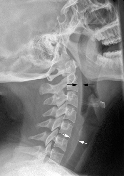
Normal soft tissue (<6mm at C2, <22mm at C6)

A study in the Jan 2011 Journal of Trauma outlined the dysphagia problem seen with placement of a halo vest. They studied a series of 79 of their patients who were treated with a halo. A full 66% had problems with their swallowing evaluation. This problem was associated with a significantly longer ICU stay and a somewhat longer overall hospital stay.

Bottom line: Suspect dysphagia in all patients with cervical fractures, especially the elderly. We don’t use halo vests very often any more, but cervical collars can exacerbate the problem by keeping the neck in an unaccustomed position. Carry out a formal swallowing evaluation, and adjust the collar (or halo) if appropriate.

Reference: Swallowing dysfunction in trauma patients with cervical spine fractures treated with halo-vest fixation. J Trauma 70(1):46-50, 2011.

Best of EAST #3: Spine MRI Usage After EAST Guidelines

In 2015, EAST published their practice guidelines for spine clearance in the obtunded blunt trauma patient. Click here to view them. They stated that a high-quality CT scan can be used to remove (clear) the cervical collar in these patients. This avoids the use of the expensive and personnel-intensive MRI clearance.

The group at UCSF used the NTDB to review the use of MRI in such patients over an 11 year period. They focused on comatose patients (GCS < 8) with an AIS head > 3 and intubation for more than 72 hours. They used logistic regression to equalize confounders while examining the use of MRI over time, before and after the guidelines were published.

Here are the factoids:

  • More than 75,000 patients from 530 trauma centers were included
  • Patients who were older, Hispanic, uninsured, or involved in a car crash were less likely to undergo spinal MRI
  • Level I centers were more likely to use MRI for clearance than Level II centers
  • Patients evaluated after release of the practice guidelines were 1.7x more likely to undergo MRI for spine clearance (!!)

The authors concluded that spinal MRI use has been increasing since 2007 despite publication of the EAST guideline.

My comments: To me, this indicates one of the following:

  1. Nobody reads the EAST guidelines, or
  2. Trauma programs believe that they alone are able to figure out what is right, and everyone else is wrong

I suspect that it is #2. For some reason, trauma programs insist on doing it their own way despite what decent evidence shows. I think that this represents a defense mechanism to minimize the cognitive dissonance that comes with defying what is published in the literature.

I always encourage programs to borrow/steal what is already out there when crafting their own practice guidelines. Someone else has already done the work, why not take advantage of it? Typically, it’s just an excuse to continue doing things the way they’ve always been done.

This incessant reinventing the wheel becomes tiresome. And for once, I don’t have many questions or suggestions for the authors. Their evidence is pretty well laid out. 

My questions for the author / presenter are:

  1. Do you use MRI for spine clearance in your obtunded blunt trauma patients? And if so, WHY?
  2. Why do you think there are demographic and trauma center level disparities? Is it the teaching environment? Something else?

To everyone else, I say “get over yourself and read the literature!”

Reference: Assessing the e3ffect of the EAST guideline on utilization of spinal MRI in the obtunded adult blunt trauma patient over time. EAST 2021, Paper 7.

What? Still Using MRI For Cervical Spine Clearance?

Cervical spine clearance as evolved considerably over the years. First, there were five views of the spine using plain radiography. Then there were three. Then we moved to CT scan with clinical clearance. And currently, many institutions are relying only on CT.

But MRI has been used as an adjunct for quite some time. Initially, it was the tie breaker in patients who had equivocal CT findings, and for a while it was used for clearance in obtunded patients. And thanks to conflicting literature and disparate studies, the occasional usage became more frequent.

The group at Cedars-Sinai Medical Center in Los Angeles  noted that the percentage of patients undergoing MRI for cervical spine evaluation at their center slowly slowly crept up from 0.9% to 5.6% over a 10 year period. They designed a study to analyze the utility of this practice and inform their future practice.

Here are the factoids:

  • Over 9,000 patients had cervical spine CT during the 10-year study period; 513 (5.6%) were positive
  • Of the 513 CT-positive patients, 290 (56%) underwent an MRI. This showed:
    • Confirmation of the major injury in 250
    • Minor injury in 40
    • Clinically significant injury was seen in only 2 which was no surprise since they both had neurologic deficits
  • Of the 8,588 CT-negative patients, only 9 had clinically significant findings and 8 of them had neurologic deficits

Bottom line: So what have we learned here? First, MRI usage at Cedars-Sinai increased over time but was really not that useful. The main use was for imaging obtunded patients or those with an obvious neurologic deficit.

More than half of patients with positive CT scans also underwent MRI. If a major injury was seen on CT, MRI confirmed it. But if the CT findings were minor, none of the MRIs added any clinically significant findings in the absence of a neurologic deficit.

And what about MRI after negative CT? In the absence of a deficit, only one had a clinically significant finding (which only required a brace).

This study shows the wisdom of monitoring “how we do it.” There is sometimes some creepage away from what the literature shows is the best practice. The best way to remedy this is to do a good study, just like the authors did. They saw a slow change in practice, investigated it, and found that there was no good clinical reason for it. This gives the trauma program the ammunition to squelch the unwelcome behavior and return the clinicians to best practices.

Reference: Is MRI becoming the new CT for cervical spine clearance? Trends in MRI utilization at a Level I trauma center. J Tra publish ahead of print, DOI: 10.1097/TA.0000000000002752, 2020.

Best of AAST #2: Cervical Spine Clearance And Distracting Injuries

Debate has forever swirled around how to clear the cervical spine. Clear clinically? CT scan plus exam? CT only? Flexion/extension views? Distracting injury?

This last one has been problematic for a long time. What is a distracting injury? Is there a difference between lower extremity wounds vs upper chest/shoulder wounds from a distraction standpoint? Is it possible to clinically clear the cervical spine if one of these injuries exist?

Finally, a multi-institutional trial was performed that strives to answer this question. Seven Level I US trauma centers participated in this 3.5 year long study. All patients with GCS > 14 underwent a standard clinical exam regardless of whether a possible distracting injury was present. Then all underwent CT evaluation of the entire cervical spine.

Here are the factoids:

  • Distracting injuries were classified into three regions: head, torso, and extremities, but no further analysis was presented in the abstract
  • Nearly 3,000 patients were enrolled and 70% had a potential distracting injury
  • A total of 233 patients (8%) had a cervical spine injury identified by CT
  • 136 patients had a cervical injury AND distracting injury, and 14 were missed by clinical exam (10%)
  • 87 patients had a cervical injury BUT NO distracting injury, and 10 were missed by clinical exam (13%)
  • Only one injury missed by clinical exam required operation

Bottom line: This study shows the usual prevalence of cervical spine injury after blunt trauma, but adds some interesting information regarding distracting injury. Basically, clinical examination will miss about 1% of patients with a negative exam, regardless of distracting injury status. Therefore, the study suggests that clinical clearance should be attempted on all patients first, regardless of “distracting injury.”

Reference: Clearing the cervical spine for patients with distracting injuries: an AAST multi-institutional trial. Session I Paper 3, AAST 2018.

Are You Still Using MRI To Clear The Cervical Spine?

There is a fairly robust  amount of data that shows that, properly performed, the cervical spine can be cleared using a high quality CT read by a highly skilled radiologist. This is true even for obtunded patients. Pooled data suggest that the miss rate in this group is only 0.017%. And MRI is not perfect either, missing significant ligamentous injury in a small number of patients.

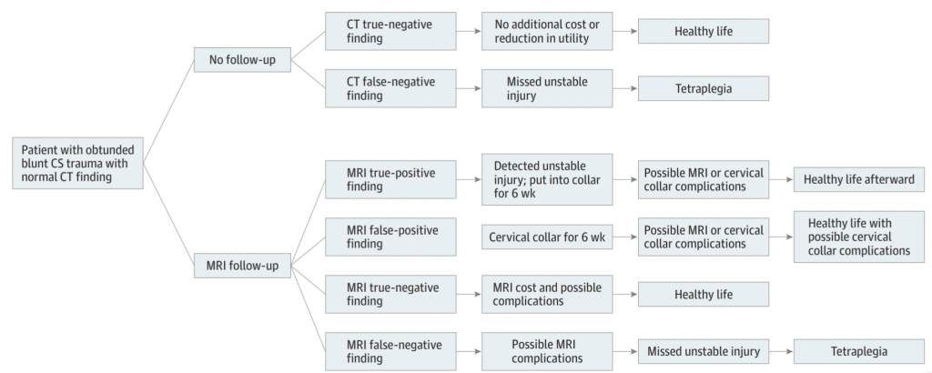
But it seems that some trauma professionals are still using MRI in some cases despite this data. The latest study on MRI focuses on the cost-effectiveness of the technique. The authors selected patients with GCS < 13 to be their obtunded group, which is probably a bit high. Nevertheless, they used a fairly sophisticated (meaning hard to understand) modeling-based decision analysis using a computerized simulation. This allowed them to compare different clearance strategies without performing large randomized clinical trials.

The authors considered MRI vs no MRI, false results, collar use and complications, MRI use with cost and complications, and the worst-case scenario of tetraplegia. Here is a flow chart of the scenarios considered. (Courtesy JAMA Surgery)

Here are the factoids:

  • The mean cost for followup vs no followup was $14K vs $1K, with no increase in quality adjusted life years (QALY)
  • No followup was the better strategy when the negative predictive value of CT was high (>98%), when the risk of an unstable injury treated with a collar turning into a permanent deficit was >25%, or if the chance of a missed injury becoming a permanent deficit was >58%
  • No followup MRI was the better strategy in all 10,000 iterations of the simulation

Bottom line: Yes, this is a fairly heavy computer simulation. But the reality is that we will never be able to design a large enough study to critically evaluate this issue and have it pass any IRB review. So it’s probably as good as it will ever get. It’s time to stop wasting money and putting obtunded patients in harm’s way by locking them into a relatively inaccessible MRI scanner for 30 minutes just to confirm the CT. Or keeping a collar until until the skin breaks down.

Here is a copy of the practice guideline we use for clearing all cervical spines, obtunded or not. Yes, there is some weirdness with soft collars, which mainly serve as a reminder to re-examine the patient at some point. But note the scan technique and requirement that it be read by a neuroradiologist for final clearance.

Related link:

Reference: Cost-effectiveness of Magnetic Resonance Imaging in Cervical
Clearance of Obtunded Blunt Trauma After a Normal
Computed Tomographic Finding