Tag Archives: Cervical spine

EAST 2018 #7: Cervical Spine Injury And Dysphagia

One of the under-appreciated complications of cervical spine fractures is dysphagia. This problem disproportionately affects the elderly, and is most common in patients with C1-C3 fractures. Swallowing becomes even more difficult when the head is held in position by a rigid cervical collar, which is the most common treatment for this injury.

How common is dysphagia in patients with cervical spine injury? What is the best way to detect it? These questions were asked by the group at MetroHealth Medical Center in Cleveland. They  retrospectively reviewed their experience with patients presenting with cervical spine injury for 14 months, then prospectively studied the use of routine, nurse-driven bedside dysphagia screening in similar patients for a year. They wanted to test the utility of screening, and judge its impact on outcome.

Here are the factoids:

  • 221 patients were prospectively studied and received a bedside dysphagia screen, but only 114 met all inclusion criteria and had the protocol properly followed (!)
  • 17% had dysphagia overall, with an incidence of 15% in cervical spine injuries and 31% in those with a concomitant spinal cord injury
  • The bedside dysphagia screen was 84% sensitive, 96% specific, with positive and negative predictive values of 80% and 97%, respectively
  • There were 6/214 patients with dysphagia complications in the retrospective group vs 0/114 in the screened group

Bottom line: This abstract actually puts a number on the incidence of dysphagia on this group of patients. I wish the patient numbers could have been higher, but they are still very good. The results are convincing, and the negative predictive value is excellent. If the screen is passed, then the patient should do well with feeds. I recommend that all patients with cervical spine injury treated with a rigid collar undergo this simple screen, and have appropriate diet adjustments to limit complications.

Here are some questions for the authors to consider before their presentation:

  • Please share the details of the nurse-driven component of the bedside dysphagia screen, and how you determine when a formal barium swallow is indicated
  • Why did your prospective study group drop from 221 to 114?
  • When did you typically perform the screen? Fracture swelling may not peak for 3 days, so early screening may not be as good as later screening.
  • This was a nice study, with a very practical and actionable result!

Reference: EAST Podium abstract #10.

A New Proposed Practice Guideline For Cervical Spine Clearance

In my last post, I reviewed a very recent prospective study on using CT scan alone for  cervical spine clearance in intoxicated patients. I believe that this is the final piece in the spine clearance puzzle to allow us to perform this task intelligently.

We’ve been accumulating more and more data that supports the use of CT scan in patients who fail clinical clearance. This failure can be due to the patient being obtunded or intoxicated, bearing a “distracting” injury, or being just plain uncooperative. Because of this, and our fear of missing a potentially devastating injury (typically because of rare anecdotal cases or urban legends), we have resorted to a significant degree of overkill. This has included, over the years, prolonged immobilization in a rigid collar, flexion/extension imaging (plain x-ray or fluoro), and MRI.

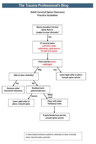
I’ve synthesized the available literature, and have drafted a simple, one sheet practice guideline for discussion. In order to use it, you must have the following:

  • A decent CT scanner – minimum 64 slice
  • A well-defined scan setup protocol – 3mm collimation, skull base to T2, 2-D reconstruction in sagittal and coronal planes (get a copy of our protocol below)
  • A skilled radiologist – neuroradiologist required

An image of the protocol can be found at the bottom of this post. I’m interested in your comments, and your comfort or discomfort with adopting something like this. Please leave comments here or on twitter.

Links: 

Reference: Cervical spine evaluation and clearance in the intoxicated patient: A prospective Western Trauma Association Multi-Institutional Trial and Survey. J Trauma 83(6):1032-1040, 2017.

It’s Time To Simplify Cervical Spine Clearance!

Cervical spine clearance is another one of those tasks that everyone seems to do their own way. Most trauma centers have an algorithm for clearance, or even two, like my center. But anytime different clinicians or centers do the same thing in different ways, it means we don’t really know what we’re doing. 

It basically means that the hard data is not there to dictate what we truly should do. So there are two alternatives:

  1. Wait for good data to become available. Unfortunately, this can take forever.
  2. Extrapolate from any existing data, and fill in the gaps with our clinical experience to come up with something that works and causes no harm.

The protocols in use at Regions Hospital are based on #2, and have been in place for over a decade. But now, we have a good example of #1 to work with.

Fortunately for us, cervical spine clearance has been evolving for decades. And as technology has improved, so has our ability to miss fewer and fewer “significant” injuries. A multi-center trial published this month provides one of the final puzzle pieces to help us settle upon a uniform cervical spine clearance guideline. It was a prospective look at intoxicated patients after blunt trauma, who can’t always participate in the process of clinical cervical spine clearance.

This three year study took place at 17 centers and specifically looked at the combination of clinical and radiographic clearance in alcohol and drug intoxicated patients. Over 10,000 patients participated in the study. There are some limitations, of course, when so many centers participate. But the pros massively outweigh the cons.

Here are the factoids:

  • The overall incidence of cervical spine injury was 10.6% (!)
  • 30% of patients were intoxicated (19% etoh, 6% drugs, 5% both (also !)
  • Intoxicated patients had a significantly lower incidence of cervical injury (8% vs 12%). (Don’t get any ideas about the old adage about being relaxed when they crash. This probably represents lower speeds involved.)
  • For intoxicated patients, sensitivity of CT scan was 94%, specificity was 99.5%, and the negative predictive value (NPV) was 99.5%
  • The NPV for clinically significant injuries in intoxicated patients was 99.9%, and no unstable injuries were missed by CT  (100% NPV) (!!)
  • When CT was negative, being intoxicated led to longer time in a collar (8 hrs vs 2 hrs)

Bottom line: Fear of clearing the cervical spine without a clinical exam, or in obtunded or intoxicated patients, is primarily due to old anecdotal reports. And much of it is not first-hand experience, but rumors of others’. What is finally becoming clear is that it is okay to clear based upon radiographic findings alone. 

Tomorrow, I’ll provide my version of a new, unified clearance protocol based on this work.

Reference: Cervical spine evaluation and clearance in the intoxicated patient: A prospective Western Trauma Association Multi-Institutional Trial and Survey. J Trauma 83(6):1032-1040, 2017.

Is Applying Or Removing That Cervical Collar Dangerous?

Cervical collars are applied to blunt trauma patients all the time. Maybe a bit too often. And most of the time, the neck is fine. It’s just those few patients that have fracture or ligamentous injury that really, truly need it.

I’ve previously written about how good some of the various types of immobilization are at limiting movement (click here). But what happens when you are actually putting them on or taking them off? Could there be dangerous amounts of movement then?

Several orthopaedics departments studied this issue using an electromagnetic motion detector on “fresh, lightly embalmed cadavers” (!) to determine how much movement occurred when applying and removing 1- and 2-piece collars. Specifically, they used an Aspen 2-piece collar, and an Ambu 1-piece. They were able to measure flexion/extension, rotation and lateral bending.

Here are the factoids:

  • There were no significant differences in rotation (2 degrees) and lateral bending (3 degrees) when applying either collar type or removing them (both about 1 degree)
  • There was a significant difference (of 0.8 degrees) in flexion/extension between the two types (2-piece flexed more). Really? 0.8 degrees is significant? Not clinically!
  • Movement was similarly small and not significantly different in either collar when removing them

Bottom line: Movement in any plane is less than 3-4 degrees with either a 1-piece or 2-piece collar. This is probably not clinically significant at all. Just look at my related post below, which showed that once your patient is in the rigid collar, they can still flex (8 degrees), rotate (2 degrees) and move laterally (18 degrees) quite a bit! So be careful when using any collar, but don’t worry about doing damage if you use it correctly.

Related post:

Reference: Motion generated in the unstable cervical spine during the application and removal of cervical immobilization collars. J Trauma 72(6):1609-1613, 2012.

Do We Need Cervical MRI Scans If The CT Is Negative?

The debate on how to clear the cervical spine just never ends. We have finally come to some degree of agreement that certain patients (awake, alert, not impaired or head injured, without distracting injury) can undergo clinical clearance alone.

But if those criteria are not met, what next? Universally, adults receive a CT scan of the cervical spine. In the majority of centers, this is coupled with a good clinical examination. And if both are negative, the collar can be removed.

But recent literature suggest that a good, high-quality cervical CT read by a skilled neuroradiologist may be good enough. This has been demonstrated in several papers involving patients who are comatose or other-wise unable to participate with a clinical exam.

Many centers and trauma professionals are still reluctant to remove the cervical collar without that clinical examination. A new study asked the question: would an MRI provide additional, significant information over and above the CT scan in those patients who could not be examined or had persistent neck pain?

A consortium of 8 Level I and II trauma centers in New England participated in this study coordinated by Yale. Blunt trauma patients who underwent MRI after negative cervical CT were considered for the study. On further review, if they received the scan because they could not be clinically evaluated, or if they had complaints of persistent neck pain, they were enrolled. CT scanners with at least 64-slice capabilities were required. There was no mention of the qualifications or special experience of the radiologists reading the images at each center.

Here are the factoids:

  • 767 patients were enrolled in this 30-month study. A total of 43% were for persistent neck pain, 44% for inability to examine, and 9% for both.
  • Nearly a quarter had an abnormal MRI scan:
    • 17% ligamentous injury
    • 4% soft tissue swelling
    • 1% disk injury
    • 1% dural hematoma
  • The collar was removed in most (88%) patients with a normal MRI, but in only 13% with ab-normal MRI
  • 11 patients underwent a surgical procedure and half had neurologic signs or symptoms. 10 of them had ligamentous injury, 1 had dural hematoma, and 1 had both

Bottom line: Looks almost compelling, right? One would think that we had better get an MRI on all of these patients! But read more closely, please. Yes, injuries were found. But did they really “require” an intervention? For some injuries, it’s a chip shot. A three column ligamentous injury equals stabilization in any textbook. But management of lesser injuries is less clear. And could some of these injuries have been recognized by a skilled neuroradiologist reading the CT image?

So what to do? There is not enough data for a universal protocol yet. Unfortunately, you will need to develop your own institutional policy based on the experience and opinions of your spine and neurosurgeons. They are the ones who will have to deal with the decision making during and after these studies. Until the definitive study comes along.

Reference: Cervical spine MRI in patients with negative CT: A prospective, multicenter study of the Research Consortium of New England Centers for Trauma (ReCONECT). J Trauma 82(2):263-269, 2017.