Tag Archives: Cervical spine

Do We Need Cervical MRI Scans If The CT Is Negative?

The debate on how to clear the cervical spine just never ends. We have finally come to some degree of agreement that certain patients (awake, alert, not impaired or head injured, without distracting injury) can undergo clinical clearance alone.

But if those criteria are not met, what next? Universally, adults receive a CT scan of the cervical spine. In the majority of centers, this is coupled with a good clinical examination. And if both are negative, the collar can be removed.

But recent literature suggest that a good, high-quality cervical CT read by a skilled neuroradiologist may be good enough. This has been demonstrated in several papers involving patients who are comatose or other-wise unable to participate with a clinical exam.

Many centers and trauma professionals are still reluctant to remove the cervical collar without that clinical examination. A new study asked the question: would an MRI provide additional, significant information over and above the CT scan in those patients who could not be examined or had persistent neck pain?

A consortium of 8 Level I and II trauma centers in New England participated in this study coordinated by Yale. Blunt trauma patients who underwent MRI after negative cervical CT were considered for the study. On further review, if they received the scan because they could not be clinically evaluated, or if they had complaints of persistent neck pain, they were enrolled. CT scanners with at least 64-slice capabilities were required. There was no mention of the qualifications or special experience of the radiologists reading the images at each center.

Here are the factoids:

  • 767 patients were enrolled in this 30-month study. A total of 43% were for persistent neck pain, 44% for inability to examine, and 9% for both.
  • Nearly a quarter had an abnormal MRI scan:
    • 17% ligamentous injury
    • 4% soft tissue swelling
    • 1% disk injury
    • 1% dural hematoma
  • The collar was removed in most (88%) patients with a normal MRI, but in only 13% with ab-normal MRI
  • 11 patients underwent a surgical procedure and half had neurologic signs or symptoms. 10 of them had ligamentous injury, 1 had dural hematoma, and 1 had both

Bottom line: Looks almost compelling, right? One would think that we had better get an MRI on all of these patients! But read more closely, please. Yes, injuries were found. But did they really “require” an intervention? For some injuries, it’s a chip shot. A three column ligamentous injury equals stabilization in any textbook. But management of lesser injuries is less clear. And could some of these injuries have been recognized by a skilled neuroradiologist reading the CT image?

So what to do? There is not enough data for a universal protocol yet. Unfortunately, you will need to develop your own institutional policy based on the experience and opinions of your spine and neurosurgeons. They are the ones who will have to deal with the decision making during and after these studies. Until the definitive study comes along.

Reference: Cervical spine MRI in patients with negative CT: A prospective, multicenter study of the Research Consortium of New England Centers for Trauma (ReCONECT). J Trauma 82(2):263-269, 2017.

EAST 2017 #3: My Neck Is Broken And It Doesn’t Hurt?

Clinical clearance of the cervical spine is a standard of care. It is usually the first method to determine if there might be an injury in patients who are awake, cooperative, and don’t have other painful distracting injuries. But appreciation of pain may be different in elderly patients, and they will frequently not notice pain from some injuries. Could this possibly impact clearance of the cervical spine?

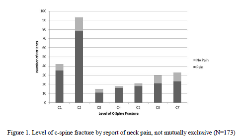
A group at Iowa Methodist performed a retrospective review of patients > 55 with diagnosed cervical spine fractures over a four year period. They were considered to have an asymptomatic injury if they did not complain of pain, or of tenderness to palpation.

Here are the factoids:

  • A total of 173 elderly patients presented with a cervical spine injury during the study period
  • 38 of them (22%) were asymptomatic
  • The asymptomatic patients tended to have higher injury severity (ISS 15 vs 10), have a significant injury in another body region (71% vs 47%), and stayed in the hospital longer (7 days vs 5)
  • A third of patients had multiple cervical fractures (symptomatic or asymptomatic?)
  • C2 was the most common fracture level

Bottom line: I have witnessed this phenomenon myself. Not all of our elders perceive pain the same way younger patients do. This study shows that it is a very significant problem. Most of the previous papers and the only review I could find do not separate out the elderly when making cervical clearance recommendations. We will probably have to develop some specific criteria to determine when a CT scan is necessary in the asymptomatic elderly patient. In the algorithm used at my hospital, age > 65 is already used to bypass clinical clearance. Looks like I’ll have to drop that to 55!

Questions and comments for the authors/presenters:

  • Since they were asymptomatic, how do you know that you didn’t miss any patients?
  • Do you have a practice guideline for cervical spine evaluation? Has it changed based on your study?
  • Be sure to break your data down by mechanism of injury for the presentation. Were there more asymptomatic patients from falls rather than car crashes? Associated fracture patterns for each mechanism?
  • What do you now recommend for clearance?
  • Suggestion: change your title to “cervical spine fractures”, not “neck fracture”.

Click here to go the the EAST 2017 page to see comments on other abstracts.

Related posts:

Reference:   Asymptomatic neck fractures: current guidelines can fail older patients. Paper #8, EAST 2017.

Cervical Spine MRI After Negative CT

dislocation-atlanto-axial-0005

There are multiple ways to clear a cervical spine! Most centers use a combination of clinical decision tools and CT scan in adults. The gold standard tie breaker, warranted or not, seems to be MRI. This tool is only used in select cases where conventional imaging is in doubt, or the clinical exam is puzzling.

Some centers clear based on CT only as long as imaging is indicated. Some use MRI in cases where patients continue to complain of midline neck pain or tenderness after negative CT. A multi-center trial encompassing 8 Level I and II centers prospectively performed MRI on patients who could not be clinically evaluated, or had persistent midline cervical pain after normal CT.

A total of 767 patients were seen over a 30 month period. Besides looking at the usual data points, the authors were interested in new diagnoses and changes in management based on the MRI results.

Here are the factoids:

  • Neck pain and inability to evaluate occurred with equal frequency, about 45%; the remaining 10% had both
  • 23% of MRIs were abnormal, with 17% ligament injury, 4% swelling, 1% disk injury, and 1% dural hematomas.
  • Patients with normal and abnormal MRI had neurologic anomalies about equally (15-19%). [Why are these patients included? Were they initially not evaluable?]
  • The cervical collar was removed in 88% of patients with normal MRI (??), and in 13% with abnormal MRI
  • After (presumably) positive MRI, 14 (2%) underwent spine surgery; 8 of these had neurologic signs or symptoms

Bottom line: I’m a bit confused. If the authors were really trying to figure out the rate of abnormal MRI after negative CT, they should have excluded the patients with known neurologic findings. These patients should nearly always have an abnormal MRI. And why did they not take the collar off of the 12% of patients with both normal CT and MRI??

Hopefully, details in the presentation next week will help explain all this. I suspect that the study will show that there are cases where CT is normal but MRI is not. The abstract does not clearly describe how many of these are clinically significant.

I admit, I’m not very comfortable clearing the cervical spine in a patient with negative CT (even if read by a neuroradiologist) and obvious midline neck pain/tenderness. I hope this study helps clarify this issue. We shall see…

Reference: Cervical spine MRI in patients with negative CT: a prospective, multicenter study of the research consortium of New England centers for trauma (ReCONECT). AAST 2016, Paper 61.

Comparison of Cervical Spine Stabilization

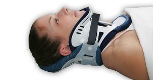
A reader recently asked what the optimal method for inline stabilization is. We’ve been pondering this question for nearly 30 years. In 1983, trauma surgeons at UCLA looked at a number of devices available at that time and tested them on normal volunteers. They measured neck motion to see which was “best.”

Here’s what they found:

  • Soft collar – In general, this decreased rotation by 8 degrees but insignificantly protected against flexion and extension. Basically, this keeps your neck warm and little else.
  • Hard collars – A variety of collars available in that era were tested. They all allowed about 8% flexion, 18% lateral movement, and 2% rotation. The Philadelphia collar allowed the least extension.
  • Sandbags and tape – Surprisingly, this was the best. It allowed no flexion and only a few percent movement in any other direction.

The Mayo clinic compared four specific hard collars in 2007 (Miami J, Miami J with Occian back, Aspen, Philadelphia). They found that the Miami J and Philadelphia collars reduced neck movement the best. The Miami J with or without the Occian back provided the best relief from pressure. The Aspen allowed more movement in all axes.

And finally, the halo vest is the gold standard. These tend to be used rarely and in very special circumstances.

Bottom line:

  • For EMS: Rigid collar per your protocol is the standard. In a pinch you can use good old tape and sandbags with excellent results.
  • For physicians: The Miami J provides the most limitation of movement. If the collar will be needed for more than a short time, consider the well-padded Occian back Miami J (see below).

References:

  • Efficacy of cervical spine immobilization methods. J Trauma 23(6):461-465, 1983.
  • Range-of-motion restriction and craniofacial tissue-interface pressure from four cervical collars. J Trauma 63(5):1120, 1126, 2007.