Tag Archives: fracture

Metal Splints – Can You X-ray Through Them?

Splinting is an important part of the trauma resuscitation process. No patient should leave your trauma resuscitation room without splinting of all major fractures. It reduces pain, bleeding, and soft tissue injury, and can keep a closed fracture from becoming an open one.

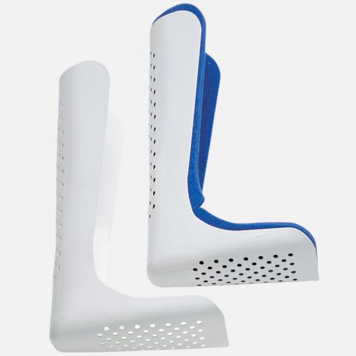
But what about imaging? Can’t the splint degrade x-rays and hamper interpretation of the fracture images? Especially those pre-formed aluminum ones with the holes in them? It’s metal, after all.

Some of my orthopedic colleagues insist that the splint be removed in the x-ray department before obtaining images. And who ends up doing it? The poor radiographic tech, who has no training in fracture immobilization and can’t provide additional pain control on their own.

But does it really make a difference? Judge for yourself. Here are some knee images with one of these splints on:

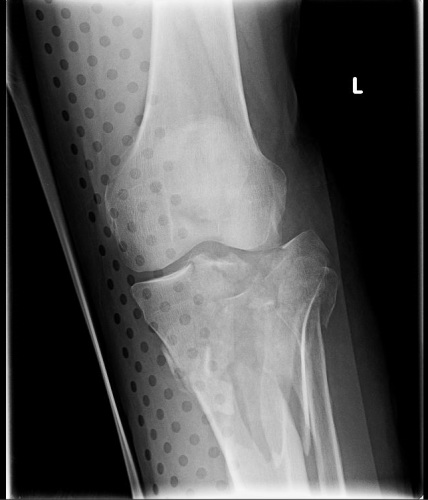
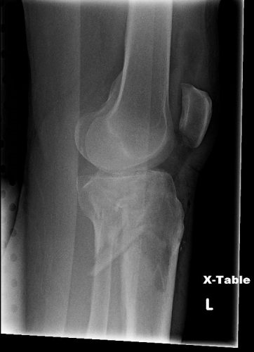
Amazingly, this thin aluminum shows up only faintly. There is minimal impact on interpretation of the tibial plateau. And on the lateral view, the splint is well posterior to bones.

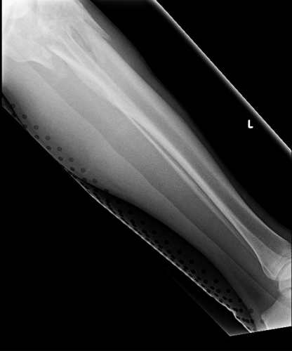
On the tib-fib above, the holes are a little distracting on the AP view, but still allow for good images to be obtained.

Bottom line: In general, splints should not be removed during the imaging process for acute trauma. For most fractures, the images obtained are more than adequate to define the injury and formulate a treatment plan. If the fracture pattern is complex, it may be helpful to temporarily remove it, but this should only be done by a physician who can ensure the fracture site is handled properly. In some cases, CT scan may be more helpful and does not require splint removal. And in all cases, the splint should also be replaced immediately at the end of the study.


EAST 2017 #3: My Neck Is Broken And It Doesn’t Hurt?

Clinical clearance of the cervical spine is a standard of care. It is usually the first method to determine if there might be an injury in patients who are awake, cooperative, and don’t have other painful distracting injuries. But appreciation of pain may be different in elderly patients, and they will frequently not notice pain from some injuries. Could this possibly impact clearance of the cervical spine?

A group at Iowa Methodist performed a retrospective review of patients > 55 with diagnosed cervical spine fractures over a four year period. They were considered to have an asymptomatic injury if they did not complain of pain, or of tenderness to palpation.

Here are the factoids:

  • A total of 173 elderly patients presented with a cervical spine injury during the study period
  • 38 of them (22%) were asymptomatic
  • The asymptomatic patients tended to have higher injury severity (ISS 15 vs 10), have a significant injury in another body region (71% vs 47%), and stayed in the hospital longer (7 days vs 5)
  • A third of patients had multiple cervical fractures (symptomatic or asymptomatic?)
  • C2 was the most common fracture level

Bottom line: I have witnessed this phenomenon myself. Not all of our elders perceive pain the same way younger patients do. This study shows that it is a very significant problem. Most of the previous papers and the only review I could find do not separate out the elderly when making cervical clearance recommendations. We will probably have to develop some specific criteria to determine when a CT scan is necessary in the asymptomatic elderly patient. In the algorithm used at my hospital, age > 65 is already used to bypass clinical clearance. Looks like I’ll have to drop that to 55!

Questions and comments for the authors/presenters:

  • Since they were asymptomatic, how do you know that you didn’t miss any patients?
  • Do you have a practice guideline for cervical spine evaluation? Has it changed based on your study?
  • Be sure to break your data down by mechanism of injury for the presentation. Were there more asymptomatic patients from falls rather than car crashes? Associated fracture patterns for each mechanism?
  • What do you now recommend for clearance?
  • Suggestion: change your title to “cervical spine fractures”, not “neck fracture”.

Click here to go the the EAST 2017 page to see comments on other abstracts.

Related posts:

Reference:   Asymptomatic neck fractures: current guidelines can fail older patients. Paper #8, EAST 2017.

Why Do They Call It: The Surgical Neck of the Humerus?

Anatomy is complex and confusing at times. Pretty much everything you can find in the human body has a name. Sometimes it makes sense. Sometimes it’s named after someone famous. And sometimes, it’s just a head-scratcher.

Let’s take the surgical neck of the humerus. Here’s an image of the proximal humerus:

proximal_humerus-14a181ca9b3646a88cc1

Notice there are two different “necks” of the humerus. You are probably familiar with the anatomic neck from your anatomy classes. But if you are a resident, an orthopedic surgeon, or someone who deals with fractures regularly, you are more familiar with the surgical neck.

The surgical neck of the humerus is the most common fracture site on the proximal humerus.  But here’s the kicker. It’s a misnomer!

Just because you see a fracture of the surgical neck of the humerus doesn’t meed it needs surgery! Indeed, many of these fractures are now successfully treated with immobilization in a sling. Your friendly neighborhood orthopedic surgeons will assess fracture stability by looking at the mechanism, exact location, involvement of the tubercles, and motion. Then they will decide on their treatment plan.

Bottom line: Don’t get suckered when someone asks you what operation is usually needed for a fracture of the surgical neck of the humerus!

Related posts: 

What You Need To Know About Frontal Sinus Fractures

Fracture of the frontal sinus is less common than other facial injuries, but can be more complex to deal with, both in the shorter and longer terms. These are generally high energy injuries, and facial impact in car crashes is the most common mechanism. Fists generally can’t cause the injury, but blunt objects like baseball bats can.

Here’s the normal anatomy:

sinus-fracture-treatment


Source: www.facialtraumamd.com

There are two “tables”, the anterior and the posterior. The anterior is covered with skin and a small amount of subcutaneous tissue. The posterior table is separated from the brain by the meninges.

Here’s an image of an open fracture involving both tables. Note the underlying pneumocephalus.

frontal_sinus1

A third of injuries violate the anterior table, and two thirds violate both. Posterior table fractures are very rare. A third of all patients will develop a CSF leak, typically from their nose.

These fractures may be (rarely) identified on physical exam if deformity and flattening is noted over the forehead. Most of the time, these patients undergo imaging for brain injury and the fracture is found incidentally. Once identified, go back and specifically look for a CSF leak. Clear fluid in the nose is, by definition, CSF. Don’t waste time on a beta-2 transferring (see below).

If a laceration is clearly visible over the fracture, or if a CSF leak was identified, notify your maxillofacial specialist immediately. If more than a little pneumocephalus is present, let your neurosurgeon know. Otherwise, your consults can wait until the next morning.

In general, these patients frequently require surgery for the fracture, either to restore cosmetic contours or to avoid mucocele formation. However, these are seldom needed urgently unless the fracture is an open fracture with contamination or there is a significant CSF leak. If in doubt, though, consult your specialist.

Related posts:

Pelvic Binder Orthosis vs Pelvic External Fixation

Yesterday, I wrote about the open book, A-P compression mechanism, pelvic fracture. In the “old” days, the recommended management for an unstable pelvis like these was application of an external fixator. In some textbooks, it was even suggested that this should be done (by orthopedics) in the resuscitation room. High volume trauma centers with ortho residents could actually pull this off, but not many others.

As the idea of pelvic orthotic binders caught on (T-POD, sheeting, etc) and was adopted by prehospital providers, and then trauma teams, the use of initial external fixation dropped off. But the idea that external fixation was the most desirable or most effective lingered on. A study from Memphis finally sheds some light on the answer to this question.

image

A 10 year retrospective review was carried out on patients presenting with multiple or severe pelvic ring fractures who had early stabilization of the pelvis. Stabilization consisted of external fixation early on, and gradually shifted to pelvic orthotic devices over the study period. They ultimately analyzed outcomes for 93 patients in each treatment group.

The authors found that transfusion needs were dramatically reduced with the orthotic devices (5 units vs 17 units at 24 hours) compared to the orthotics. About a quarter of patients in each group went to angiography, and even in those patients the transfusion need remained lower in the orthotic device group. Hospital length of stay was also significantly shorter in this group (17 vs 24 days). There was no difference in mortality.

Bottom line: Although this is a small, retrospective study it easily showed significant results and will probably never be repeated. Use of a pelvic orthotic device (POD) resulted in less blood replacement and shorter stays in hospital. This technique is simple, cheap and quick, an ideal combination. But does a sheet count as an orthotic device? We don’t know. It’s really cheap, but probably a bit less controlled than a POD. If you have a real POD in your ED or your ambulance, use it. If not, apply the sheet, which will be described tomorrow with other binders.

Tomorrow: what’s the “best” pelvic binder?

Reference: Emergent pelvic fixation in patients with exsanguinating pelvic fractures. JACS 204:935-942, 2007.