Galleries

Hypothermia And Wound Infection In Trauma

For the most part, hypothermia is a bad thing for trauma patients. Its impact on bleeding and mortality has long been known. A paper just out now implicates it in surgical site infections as well. This fact has already been shown for some types of elective surgery (colorectal), but it appears to be a factor in trauma laparotomy as well.

A retrospective review of 524 patients who underwent a trauma lap looked at the correlation of surgical site infection (SSI) and the depth and duration of hypothermia. The mean low temp across all cases was 35.2° C (!). Nearly a third had at least one measurement below 35° C. About 36% of all patients developed an SSI.

  • Hypothermia is a common problem in these patients!
  • 35 C was the nadir temp most predictive of developing an infection
  • Every degree below 35 C more than tripled the risk of SSI

Bottom line: Yet one more reason to avoid hypothermia in our trauma patients! This effort begins with prehospital providers doing their best to insulate and keep patients warm. The trauma team also has a responsibility to heat up the room and keep the patient covered as much as possible. Baseline temp should be obtained in all major trauma patients. And if they do end up in the operating room, anesthesia needs to monitor the temp closely and keep the surgeon apprised of any concerning drops.

Related posts:

Reference: The Effects of Intraoperative Hypothermia on Surgical Site Infection: An Analysis of 524 Trauma Laparotomies. Annals of Surgery 255(4):789-795, 2012.

Print Friendly, PDF & Email

Prehospital To Trauma Team Handoff: A Solution

I’ve written about handoffs between EMS and the trauma team over the past two days. It’s a problem at many hospitals. So what to do?

Let’s learn from our experience in the OR. Best practice in the operating room mandates a specific time out process that involves everyone in the OR. Each participant in the operation has to stop, identify the patient, state what the proposed procedure and location is, verify that the site is marked properly, and that they have carried out their own specific responsibilities (e.g. infused the antibiotic).

Some trauma centers have initiated a similar process for their trauma team as well. Here’s how it works:

  • The patient is rolled into the resuscitation room by EMS personnel, but remains on the stretcher.
  • Any urgent cares continue, such as ventilation.
  • The trauma team leader is identified and the EMS lead gives a brief report while everyone in the room listens. The report consists of only mechanism, all identified injuries, vital signs (including pupils and GCS), any treatments provided. This should take no more than 30 seconds.
  • An opportunity for questions to be asked and answered is presented
  • The patient is moved onto the hospital bed and evaluation and treatment proceed as usual.
  • EMS personnel provide any additional information to the scribe, and may be available to answer any additional questions for a brief period of time.

Bottom line: This is an excellent way to improve the relationship between prehospital and trauma team while improving patient care. It should help increase the amount of clinically relevant information exchanged between care providers. Obviously, there will be certain cases where such a clean process is not possible (e.g. CPR in progress). I recommend that all trauma programs consider implementing this “Trauma Activation Time Out For EMS” concept.


Related posts:

Print Friendly, PDF & Email

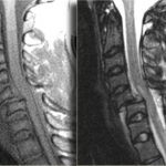
Clearing The Cervical Spine With MRI

If you follow the trauma literature, clearance of the cervical spine in obtunded patients is confusing at best. Although there is some literature out there that suggests that a good cervical CT alone is adequate, I’m not a believer. I’ve seen a case where the radiologist called the scan normal and a good spine surgeon called an injury and was right. So I’m reluctant to use CT alone because the skills of radiologists vary widely. I might be able to believe a dedicated neuroradiologist, but you can’t guarantee one will be reading your patient’s images.

So I fall back on the routine of clearing the bones with a CT scan, and the ligaments with something else. That something else could be a clinical exam (not available in the obtunded patient), flexion-extension images under fluoroscopy (makes a lot of people nervous), keeping the patient in a collar for weeks (skin breakdown), or an MRI. The problem is that there is little guidance in the literature regarding how good MRI is or the best way to use it.

A recent paper in the Journal of Trauma retrospectively looked at 512 out of 17,000 patients (!) seen over 5 years at one trauma center who had both CT and MRI of the c-spine. They wanted to determine if MRI was of any value in cervical spine clearance. Only 150 met the inclusion criteria (GCS<13, no obvious neuro deficit, normal CT). Half of the MRIs were normal. Of the abnormal ones, 81% showed a ligamentous or soft tissue injury. None were deemed unstable and no specific management was needed for any of the abnormal scans.

The authors interpreted their data as showing that MRI provided no additional useful information. However, numbers were (very) small, so the likelihood of them seeing someone with an unstable ligamentous injury was low. Could it be that they showed that MRI detected stable injuries well, and that they could essentially remove the collar based on that?

Bottom line: We still don’t know how to use MRI for clearance. My bias (no good data I can find) is that it is good in suggesting ligamentous injury via nearby edema. If this injury involves only one set of ligaments, it is very likely a stable one and the collar can be removed. If it involves several groups of ligaments, that is probably not the case. And how soon do we have to get the MRI after injury? Some have suggested that 72 hours is the ideal window because edema decreases afterwards. Sounds reasonable, but I can’t find a shred of evidence in the literature. For now, I’ll get an MRI within 72 hours and if it is abnormal, pass the buck to my neurosurgical colleagues so they can gnash their teeth, too.

I would be very happy if someone can help me out and point me towards some good literature on this topic!

Reference: The value of cervical magnetic resonance imaging in the evaluation of the obtunded or comatose patient with cervical trauma, no other abnormal neurological findings, and a normal cervical computed tomography. J Trauma 72(3):699-702, 2012.

Print Friendly, PDF & Email

Why Do They Call Them Rounds?

Face it. Everyone uses this term. But where did it come from? After a little digging, I think I’ve found the answer. I’m sure someone will step forward and offer another explanation, but the origins of some of our traditions grow foggy with time.

Supposedly, the term “rounds” was introduced by Sir William Osler, the famed physician, while he was at Johns Hopkins hospital in Baltimore. The original building was built in the 1880s and had a round dome. Osler and his trainees had to walk circular hallways to see their patients. I’ve not been there, but looking at the picture above, the corners of the building appear to be octagonal patient wards as well.

The term has stuck with us, and today just about every discipline from prehospital to rehab medicine use it! If anybody has another theory or correction, please let me know!

Reference: CIRCULAR HOSPITAL WARDS: PROFESSOR JOHN MARSHALL’S CONCEPT AND ITS EXPLORATION BY THE ARCHITECTURAL PROFESSION IN THE 1880s. Medical History 32:426-448, 1988.

Print Friendly, PDF & Email

Figure It Out! The Answer

Several readers emailed the correct answer yesterday. The picture is a child with a bicycle handlebar injury to the epigastrium. The plastic grip over the end of the handlebar has a small hole in the middle leaving the distinctive mark seen in the photo.

Children are more likely to sustain significant injuries from this mechanism because they have little muscle in their abdominal wall, so it can’t protect as well as it does in adults. Everything between the handlebar and the spine gets crushed together, frequently resulting in serious injury.

Possible injuries include:

  • Pancreatic injury / transection
  • Liver laceration (left lobe)
  • Duodenal injury / hematoma
  • Retrohepatic vena cava injury

I’ve listed them in what I believe to be the usual order. The literature varies a bit because there aren’t a lot of series published. In this case, the injury was a pancreatic transection.

Bottom line: Handlebar injuries in children (and to a lesser degree, adults) are a significant marker for serious abdominal injury. CT scan is mandatory to find the diagnosis. Proper management of a pancreatic injury is a good topic for a future post!

Print Friendly, PDF & Email