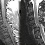
Clearing The Cervical Spine With MRI
If you follow the trauma literature, clearance of the cervical spine in obtunded patients is confusing at best. Although there is some literature out there that suggests that a good cervical CT alone is adequate, I’m not a believer. I’ve seen a case where the radiologist called the scan normal and a good spine surgeon called an injury and was right. So I’m reluctant to use CT alone because the skills of radiologists vary widely. I might be able to believe a dedicated neuroradiologist, but you can’t guarantee one will be reading your patient’s images.
So I fall back on the routine of clearing the bones with a CT scan, and the ligaments with something else. That something else could be a clinical exam (not available in the obtunded patient), flexion-extension images under fluoroscopy (makes a lot of people nervous), keeping the patient in a collar for weeks (skin breakdown), or an MRI. The problem is that there is little guidance in the literature regarding how good MRI is or the best way to use it.
A recent paper in the Journal of Trauma retrospectively looked at 512 out of 17,000 patients (!) seen over 5 years at one trauma center who had both CT and MRI of the c-spine. They wanted to determine if MRI was of any value in cervical spine clearance. Only 150 met the inclusion criteria (GCS<13, no obvious neuro deficit, normal CT). Half of the MRIs were normal. Of the abnormal ones, 81% showed a ligamentous or soft tissue injury. None were deemed unstable and no specific management was needed for any of the abnormal scans.
The authors interpreted their data as showing that MRI provided no additional useful information. However, numbers were (very) small, so the likelihood of them seeing someone with an unstable ligamentous injury was low. Could it be that they showed that MRI detected stable injuries well, and that they could essentially remove the collar based on that?
Bottom line: We still don’t know how to use MRI for clearance. My bias (no good data I can find) is that it is good in suggesting ligamentous injury via nearby edema. If this injury involves only one set of ligaments, it is very likely a stable one and the collar can be removed. If it involves several groups of ligaments, that is probably not the case. And how soon do we have to get the MRI after injury? Some have suggested that 72 hours is the ideal window because edema decreases afterwards. Sounds reasonable, but I can’t find a shred of evidence in the literature. For now, I’ll get an MRI within 72 hours and if it is abnormal, pass the buck to my neurosurgical colleagues so they can gnash their teeth, too.
I would be very happy if someone can help me out and point me towards some good literature on this topic!
Reference: The value of cervical magnetic resonance imaging in the evaluation of the obtunded or comatose patient with cervical trauma, no other abnormal neurological findings, and a normal cervical computed tomography. J Trauma 72(3):699-702, 2012.


