Galleries

New Technology: The End Of Handwashing?

All healthcare professionals are notoriously bad about washing their hands, especially doctors. A variety of things have been developed to help us keep our hands clean, including simple soap and water, barriers like gloves, and various gels and foams (which I swear I can taste in my mouth 10 minutes later, even though I’m pretty sure I’m not putting my fingers there).

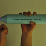
A recently published paper from China is shining new light on this topic (get it?). Researchers developed a hand-held, battery-powered plasma flashlight that gets rid of bacteria on skin in a flash. It costs less than $100 to produce and runs on a 12V battery. 

This device was found to inactivate all bacteria in a 17-layer biofilm containing a very hardy organism, enterococcus faecalis. It does not produce UV radiation, and the exact mechanism for the bacterocidal effect is unclear. There was no adverse effect on skin.

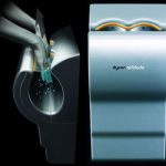
The main drawbacks to this device are that it only produces a small area of plasma, and it takes 5 minutes to kill all the bacteria. But take this to the next logical step. Many of you are familiar with the Dyson Airblade hand dryers found at many airports. Suppose you could produce a more intense plasma field using a more robust power supply (power line or ambulance power system) in a device that you could just pass your hands through to disinfect them? 

And if you really want to improve compliance, hook the unit to the door control so the doctor can’t even walk into a patient room without passing his hands through it!

Reference: Inactivation of a 25.5µm Enterococcus faecalis biofilm by a room-temperature, battery-operated, handheld air plasma jet. Journal of Physics D: Applied Physics 45(2012):165205 (5p), 2012.

Print Friendly, PDF & Email

Best Of: Flexion / Extension Views of the Cervical Spine

I’ve gotten a number of requests about the use of flexion-extension views of the cervical spine to aid in spine clearance. Here are some answers to common questions about this practice.

Clearance of the cervical spine can often be done using clinical criteria alone (see this video at http://youtu.be/NhjF9kDOcjE). If this is not possible, a combination of radiologic and clinical evaluation is usually carried out. 

In some cases, radiographic studies (usually CT) are normal, but there is pain on clinical exam. Our next step is to send the patient to xray for flexion and extension views. This exam is performed by removing the collar while the patient is sitting, so the thoracic and lumbar spines must be clear before ordering this. The patient then gently flexes and extends the neck to their limits of comfort. Images are then obtained at the limits of flexion and extension. The premise is that a normal, awake patient cannot and will not move their neck beyond their comfort level to the point where they could cause themselves neurologic injury. 

It is very important that you look at the images yourself. The radiologist may review the images and will report that “there is no evidence of subluxation at the limits of flexion and extension.” But the patient may have barely moved their neck!

The question is: how much flexion and extension do you need to have to clear the spine?

The answer is not easy to find, and is buried in literature from the 1980s and 90s. According to the EAST guidelines, the ideal amount is 30 degrees from neutral for both flexion and extension. This is not always achievable in elderly patients, so in those cases you must use your judgment. Talk to the patient to find out if they stopped moving their neck forward or backward due to pain, or because they just can’t move it that far.

Trouble signs to look for are:

  • Subluxation of more that 2mm at any level
  • Angulation of more than 11 degrees

Any abnormality should prompt a spine consult.

If the study is not abnormal but the amount of flexion and/or extension is not adequate, there are two options. First, just leave the collar in place and try again in a week or so and try again. This will allow any soft tissue injuries to get better and may allow a successful repeat study. The alternative is a more costly and less well-tolerated MRI.

Related posts:

References:

  1. EAST Practice Guidelines, Identifying Cervical Spine Injuries Following Trauma – Update (2000).
  2. Defining radiographic criteria for flexion-extension studies of the cervical spine. Robert Knopp et al. Ann Emerg Med. 2001 Jul;38(1):31-5.
Image: C5-6 subluxation with only a slight amount of flexion
Print Friendly, PDF & Email

Best Of: Enoxaparin And Pregnancy

Pregnant women get seriously injured, too. And pregnancy is an independent risk factor for deep venous thrombosis. We reflexively start at-risk patients on prophylactic agents for DVT, the most common being enoxaparin. But is it safe to give enoxaparin during pregnancy?

Studies have looked at drug levels in cord blood when the mother is receiving enoxaparin, and none has been found. No specific bleeding complications have been identified, either. So from the baby’s standpoint, administration is probably safe.

However, there are two other issues to consider. In a study looking at the use of enoxaparin for prophylaxis in women with a mechanical heart valve, 2 of 8 women (and their babies) died. Both suffered from clots that developed and blocked the valves. Most likely, the standard dose of enoxaparin was insufficient, so monitoring of anti-Factor Xa levels must be done.

The other problem lies in the multi-dose vial of Lovenox (Sanofi-Aventis). Each 100mg vial contains 45mg of benzyl alcohol, which has been associated with a fatal “gasping syndrome” in premature infants. The individual dose syringes do not have this preservative.

Bottom line: It is probably safe to give enoxaparin to pregnant women after trauma. However, it is unclear if the dose needs to be increased to achieve adequate prophylaxis. Only consider using this medication after consultation with the patient’s obstetrician, and use only the individual dose syringes. Otherwise fall back to standard subcutaneous non-fractionated heparin (even though it is a Category C drug by FDA; it is still considered the anticoagulant of choice during pregnancy).

Print Friendly, PDF & Email

Do You Really Need To Repeat That Xray?

It happens all the time. You get that initial chest and/or pelvic xray in the resuscitation room while evaluating a blunt trauma patient. A few minutes later the tech returns with another armful of xray plates to repeat them. Why? The patient was not centered properly and part of the image is clipped.

Do you really need to go through the process of setting up again, moving the xray unit in, watching people run out of the room (if they are not wearing lead, and see my post below about how much radiation they are really exposed to), and shooting another image? The answer to the question lies in what you are looking for. Let’s address the two most common (and really the only necessary) images needed during early resuscitation of blunt trauma.

First, the chest xray. You are really looking for 3 things:

  • Big air (pneumothorax)
  • Big blood (hemothorax)
  • Big mediastinum (hinting at aortic injury)

Look at the clipped xray above. A portion of the left chest wall is off the image. If there were a large pneumothorax on the left, would you be able to see it? What about a large hemothorax? And the mediastinum is fully included, so no problem there. So in this case, no need to repeat immediately.

The same thing goes for the pelvis. You are looking for gross disruption of the pelvic ring, especially posteriorly because this will cause you to intervene in the ED (order blood, consider wrapping the pelvis). So if parts of the edges or top and bottom are clipped, no big deal.

Bottom line: Don’t let the xray tech disrupt the team again by reflexively repeating images that are not technically perfect. See if you can use what you already have.  And how do you decide if you need to repeat it later, if at all? Consider the mechanism of injury and the physical exam. Then ask yourself if there is anything you could possibly see that was not imaged the first time that would change your management in any way. If not, you don’t need it. But it certainly will irritate the radiologists!

Related posts: 

Print Friendly, PDF & Email

Diffuse Axonal Injury (DAI)

Trauma professionals tend to focus on the two extremes of TBI: mild concussive injury because we see so much of it, and very severe injury that we have to work so hard to keep the patient alive. Today I’ll write about the one in the middle, diffuse axonal injury (DAI). People don’t talk about it nearly as much, and it seems kind of mysterious.

DAI is also known as a shear injury, because that’s what we think happens to the brain at the time of injury. Officially, it is diagnosed when a patient remains in a coma for more than 6 hours without a demonstrable mass lesion from bleeding seen on CT. It is seen in about 15% of trauma ICU patients with head injury. Essentially the substance of the brain moves around enough to disrupt a critical mass of axonal connections that results in prolonged unconsciousness. It then takes time to try to rebuild those connections and restore consciousness and some degree of cognition. Mechanisms which result in sudden acceleration or deceleration of the brain may cause this condition, and rotational forces which spin the head suddenly seem to be even worse.

CT scan of the head frequently shows no unusual findings. On occasion, small punctate hemorrhages may be seen. These are generally bad prognostic signs, because CT is so much less sensitive in showing these compared to MRI. Here are some key points about DAI:

  • If the head CT is negative,and all recreational drugs have worn off and the patient still doesn’t wake up, DAI is likely.
  • MRI can confirm the diagnosis, but is not good for giving a prognosis
  • Slow recovery of consciousness or failure to recover correlates with death
  • Hyperglycemia and the presence of a subdural also are highly correlated with mortality

Bottom line: The diagnosis of DAI can generally be made clinically with the assistance of head CT. MRI is not very useful, unless it is needed to confirm the diagnosis. It does not predict speed or degree of recovery so is otherwise not very useful. Supportive care, avoidance of complications and early therapy and rehab are the best treatments we have to offer.

Reference: Diffuse axonal injury in patients with head injuries: an epidemiologic and prognosis study of 124 cases. J Trauma 71(4):838-846, 2011.

Print Friendly, PDF & Email