Galleries

Simple Tracheobronchial Injury


Injury to the airway has the potential to be a catastrophe, with rapid deterioration and death. Occasionally the injury is less dramatic with a slow air leak, but it can still present a diagnostic and management challenge. 

These lower airway injuries can occur after either blunt or penetrating trauma. The penetrating ones are relatively simple to diagnose because the wound tract is known and, if stable, a trip to CT demonstrates the problem area. 

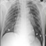
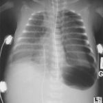
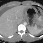
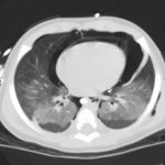
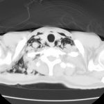
Blunt lower airway injury is a bit trickier. These typically require a high energy mechanism, such as a motor vehicle crash. Up to half of these injuries are not diagnosed immediately. Typically, unexpected air on the chest xray is identified. Less commonly, subcutaneous emphysema appears and prompts more investigation.

Previously, the gold standard for diagnosis was bronchoscopy. CT has gotten so good that even smaller bronchial injuries can be identified, so CT is now the diagnostic study of choice. Management of injuries that do not threaten the airway consists of close observation. Smaller ones may heal on their own without complication. Larger injuries usually continue to leak and do not heal. If ventilation problems develop, either from persistent large pneumothorax or large amounts of air dissecting into the neck, intubation will be required. However, positive pressure may exacerbate the problem, so low pressure ventilation modalities must be used. A prompt trip to the OR will be required in such cases.

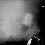
Bottom line: Simple (slow leak) tracheobronchial injuries are uncommon, but are seen after major blunt trauma and any kind of thoracic penetrating injury. The best way to diagnose the exact problem and location is thin cut CT of the chest. Injuries with minor clinical dysfunction can be admitted to the trauma service and observed. If the leak does not resolve, or causes breathing problems, a thoracic surgeon will very likely need to intervene to repair the problem.

Related posts:

Print Friendly, PDF & Email

The Deep Sulcus Sign

Pneumothorax is frequently difficult to diagnose in the resuscitation room. Sometimes it is obvious, with a hypoxic patient and absent breath sounds. But not usually. Most of the time we rely on a chest xray to help make the diagnosis.

Unfortunately, the good old chest xray only shows a pneumothorax about 30-50% of the time. A big part of the problem is that our patients are usually supine to protect their spine. A small pneumothorax make float anteriorly in the supine position, and if it is not big enough to wrap around the lateral edge of the lung, it may remain invisible. So you need to look for gross and subtle signs on the image that will help make the diagnosis. The deep sulcus sign is one of the more subtle signs. 

Simply stated, the deep sulcus sign is a radiolucent (dark) lateral sulcus where the chest wall meets the diaphragm. The amount of lung in this area is less, so a small amount of air will tend to darken the area making it more prominent. Look at patient left in the left photo, and compare to their right side. It is much darker and appears to extend lower than usual. In more extreme cases, the amount of air just above the diaphragm may make it appear inverted (right photo).

Bottom line: If you see a deep sulcus sign on the chest xray image, strongly consider pneumothorax. If the patient begins to have hemodynamic problems, needle the chest and chase with a chest tube. If they remain stable, the patient will still require a chest tube. Chest xray always underestimates the true size of the pneumothorax. Place the usual size chest tube and manage per your usual protocol. And, as always, use your best sterile technique and definitively identify the proper side before placing the tube.

Related posts:

Print Friendly, PDF & Email

What The Heck? The Answer

The photo on Friday shows a woman who had been run over by her own car. The vehicle had rolled over her pelvis and stopped, requiring extrication. The most likely injury is an open book pelvic fracture with significant diastasis and/or bilateral unstable sacral fractures.

If you see this clinical presentation there are several things you need to do immediately:

  1. Call for blood. Losses will be large, so you may even want to consider activating your massive transfusion protocol.
  2. Call an orthopedic surgeon. External stabilization will be needed to help decrease blood loss.
  3. Consider early intubation for control of pain. You will be doing a lot, and a patient in agony will slow you down. Your patient is already hypovolemic, so plan your drug choices accordingly.
  4. Search for evidence of an open fracture. Do a good rectal and vaginal exam looking for blood.

The pelvic xray is poor quality, but shows the major problem, a 10cm pubic diastasis from the open book pelvis fracture. Wrapping the pelvis may be of some help, but consult your orthopedic surgeon first. This pelvis is probably not connected to the spine anymore, so wrapping may have variable results.

Print Friendly, PDF & Email

Delayed Splenic Rupture: Part 2

Yesterday I wrote about the history of “delayed splenic rupture.” Today I’ll discuss how to deal with it.

If possible, try to avoid ever having to mess around with this clinical problem. If you order an abdominal CT after blunt trauma and see a splenic contrast blush of either type (pseudoaneurysm or extravasation, see left photo), then deal with it before the patient even knows he has a problem. A trip to interventional radiology will usually solve the problem. And if embolized, these patients almost never come back with a bleeding problem.

As I’ve said many times before, if the patient is hemodynamically compromised, then an OR visit is required. The usual solution is splenectomy. Some recommend repairing the spleen, but this is technically more difficult than it sounds, and it is difficult for the surgeon to sleep soundly after performing one of these.

Lets say you inherited one of these from someone else, or ignored the warning signs on the initial CT. The usual time frame for presentation to the ED with acute bleeding is 7 to 10 days after the initial injury. If they are not stable, physical exam or FAST will quickly direct you to the OR, once again for splenectomy. Some patients will stabilize with fluids and can safely be sent to CT scan.

Once the CT confirms what the problem is, a trip to interventional radiology is in order if the patient remains stable. Here is the key: the radiologist must embolize something! If they find a bleeding vessel, then they can selectively embolize it. If they don’t, then the main splenic artery should be embolized. This will decrease the arterial pressure head, but won’t eliminate it. It will decrease the likelihood of additional bleeding as much as possible.

At this point, the patient should be admitted to the trauma service and monitored using your solid organ injury protocol. If they have any hemodynamic issues, it’s time to remove the spleen. Remember, this is the third time they’ve had a problem, and like in baseball, their spleen is out! Attempted splenorrhaphy at this point is pointless and may lead to yet another operation.

Related posts:

Print Friendly, PDF & Email