The Deep Sulcus Sign
Pneumothorax is frequently difficult to diagnose in the resuscitation room. Sometimes it is obvious, with a hypoxic patient and absent breath sounds. But not usually. Most of the time we rely on a chest xray to help make the diagnosis.
Unfortunately, the good old chest xray only shows a pneumothorax about 30-50% of the time. A big part of the problem is that our patients are usually supine to protect their spine. A small pneumothorax make float anteriorly in the supine position, and if it is not big enough to wrap around the lateral edge of the lung, it may remain invisible. So you need to look for gross and subtle signs on the image that will help make the diagnosis. The deep sulcus sign is one of the more subtle signs.
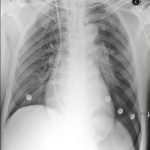
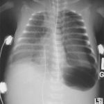
Simply stated, the deep sulcus sign is a radiolucent (dark) lateral sulcus where the chest wall meets the diaphragm. The amount of lung in this area is less, so a small amount of air will tend to darken the area making it more prominent. Look at patient left in the left photo, and compare to their right side. It is much darker and appears to extend lower than usual. In more extreme cases, the amount of air just above the diaphragm may make it appear inverted (right photo).
Bottom line: If you see a deep sulcus sign on the chest xray image, strongly consider pneumothorax. If the patient begins to have hemodynamic problems, needle the chest and chase with a chest tube. If they remain stable, the patient will still require a chest tube. Chest xray always underestimates the true size of the pneumothorax. Place the usual size chest tube and manage per your usual protocol. And, as always, use your best sterile technique and definitively identify the proper side before placing the tube.
Related posts:
