Tag Archives: technology

First, There Was REBOA, And Now… GROA?!

REBOA (Resuscitative Endovascular Balloon Occlusion of the Aorta) is a “newer” resuscitative technique that has actually been around since the Korean War. It was first used to treat two injured soldiers, and although they ultimately died, it spurred research into the technique and its applications.

Balloon occlusion techniques were then adopted by vascular surgeons and were found to be useful as an adjunct in managing ruptured abdominal aneurysms. A slow trickle of studies on human use in trauma began to surface. But when autopsy studies carried out during the Gulf War showed that uncontrolled torso hemorrhage was a major cause of death, research in the technique exploded.

First, there were a rash of pig studies evaluating the feasibility of using a percutaneously placed occlusion device in the early 2010s. This transitioned to human studies around 2014, and after that we were off to the races. Over 100 papers on REBOA are now published each year.

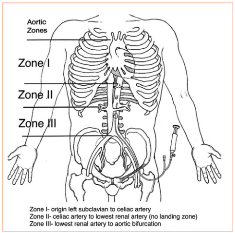
REBOA has been shown to have some advantage in patients with abdominal or pelvic sources of bleeding. The catheter is inserted in the groin and the balloon inflated in one of two zones, depending on the location of the hemorrhage (see diagram below). For abdominal bleeding, it is inserted just above the diaphragm in Zone I. For pelvic bleeding, it is inserted below the takeoff of the visceral arteries and above the aortic bifurcation, in Zone III.

In the US, REBOA catheters are only inserted upon arrival to the hospital. There are a few random reports of field placement where a physician is part of the prehospital team. By definition, this technique is generally not available in austere environments, only upon arrival in the emergency department.

Researchers at the University of Michigan began looking for an alternative technique that could be applied in the field by non-physicians. They noted the close anatomic relationship of the distal esophagus, proximal stomach, aorta, and thoracic vertebrae, and designed a device to compress the aorta against the spine in this area.

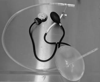
They developed a prototype device which they named GROA (gastro-esophageal resuscitative occlusion of the aorta). It consists of a gastro-esophageal tube with an ovoid balloon, an air pump with pressure measurement device, and an external compression device. Here is a picture of the device:

And here’s a diagram of what it looks like when inserted:

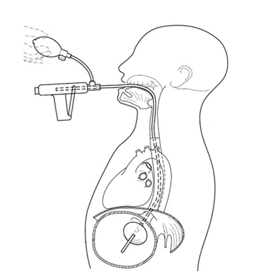
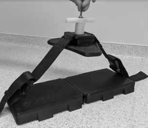
The tube is inserted and the balloon inflated. The external compression device is then placed around the patient, with the top plate located over the epigastrium and the bottom plate under the patient. It is designed to apply anterior pressure over the balloon, but to avoid circumferential constriction of the abdomen.

Bottom line: This device is an interesting development in the balloon occlusion space. As with early studies of REBOA, GROA is currently being investigated using a pig model. If it appears to be beneficial, it will still be several years before it makes the jump to human subjects. If effective, this concept would allow prehospital providers to apply some degree of hemorrhage control when it originates in the abdominal cavity.

There are currently exactly three papers on this new technique, and I have included the references below if you are interested in reading them. I’m sure there are many more to come and it may eventually be competing with REBOA for journal space.

There is one consideration to be aware of when reading these papers that is similar to much of the research on REBOA. Two of the authors have a financial interest in the company that licenses the GROA technology. And in the most recent study, another one of the authors is an advisory board member for one of the manufacturers of REBOA catheters. For these reasons, it is less likely that they will publish papers that are not favorable to the product. So read critically!

References:

  • Gastroesophageal resuscitative occlusion of the aorta: Physiologic tolerance in a swine model of hemorrhagic shock, Journal of Trauma and Acute Care Surgery: December 2020 – Volume 89 – Issue 6 – p 1114-1123 doi: 10.1097/TA.0000000000002867
  • Gastroesophageal resuscitative occlusion of the aorta prolongs survival in a lethal liver laceration model, Journal of Trauma and Acute Care Surgery: May 2022 – Volume 92 – Issue 5 – p 880-889 doi: 10.1097/TA.0000000000003444
  • Tandem use of Gastroesophageal Resuscitative Occlusion of the Aorta followed by REBOA in a Lethal Liver Laceration Model, Journal of Trauma and Acute Care Surgery: June 10, 2022 –  doi: 10.1097/TA.0000000000003719

Finding Tough-To-See Veins – Revisited

I’m always interested in technology that makes what we do easier, and this item seems to be in the news again. It’s not new technology any more; I first wrote about this way back in 2011. Here’s an objective look at an interesting machine that’s been around for a while. It uses near-infrared light to detect skin temperature changes to allow it to map out veins. It then projects an image of the map in real time onto the skin. In theory, this should make IV starts easier (as long as you can keep your head out of the way of the projector).

One of the first published papers was from Providence, Rhode Island.  It evaluated this device to see if it could simplify IV starts in a tertiary pediatric ED. It was a prospective, randomized sample of 323 children from age 0 to 17 looking at time to IV placement, number of attempts, and pain scores.

Unfortunately, the authors did not find any differences. They found that nearly 80% of IVs were started on the first attempt with or without the VeinViewer, which is less than the literature reported 2-3 attempts. This is most likely due to the level of experience of the nurses in this pediatric ED.

The authors did a planned subgroup analysis of the youngest patients (age 0-2) and found a modest decrease in IV start time (46 seconds) and the nurse’s perception of the child’s pain. Interestingly, the parents did not appreciate a difference in pain between the two groups. This may be due to the VeinViewer’s pretty green display acting as distraction therapy for the child.

The Children’s Hospital of Colorado repeated this study and reported their results earlier this year. And unfortunately they had similar findings. There were no significant differences in success rates using the VeinViewer. Also, nurses did not note any difference in their perceived insertion skills or confidence.

Bottom line: Once again, it seemed like a good idea. But that doesn’t necessarily mean that it is. And we always automatically reach for the new shiny toy. This paper points out the importance of carefully reviewing all new (read: expensive at about $20,000 each) technology before blindly implementing it. In this case, an expensive peice of equipment can’t improve upon what an experienced ED or pediatric nurse can already accomplish.


References: 

  1. VeinViewer-assisted intravenous catheter placement in a pediatric emergency department. Acad Emerg Med, 18(9):966-971, 2011.
  2. Utilization of a biomedical device (VeinViewer® ) to assist with peripheral intravenous catheter (PIV) insertion for pediatric nurses. J Spec Pediatr Nurs. 23(2):e12208, 2018.

I have no financial interest in Christie Digital Systems, distributor of the VeinViewer Vision®.

New Technology: The AED Drone

YouTube player

The media tends to give drones a bad name. And certainly, there are careless operators out there who may give drone operators a bad name. But it seems that everyone is getting in the game. Amazon wants to use drones to deliver your orders. Police use them to find missing people, and criminals. Parks use them to protect animals and property.

But how about some medical uses? Sure, they can be used to access austere environments, and potentially to deliver medical supplies. But here is an example of a very creative use. It’s an AED drone!

This drone was designed from the ground up to provide emergency assistance for cardiac arrest. It’s got audio, video, and is a flying defibrillator. Watch this 3 minute video to see how it works and how it was made.

New Technology: The Next Generation Antibiotic Bead?

A number of surgical disciplines use antibiotic beads to deliver antimicrobial drugs to sites that may not have ideal serum penetration. Unfortunately, beads require multiple operations for placement and replacement until the desired effect is achieved.

What if there was a way of delivering antimicrobial therapy directly to the tissues that works for up to two weeks, then dissolves with no trace? A system that does this is being developed by engineers at Tufts University and the University of Illinois at Urbana. They created a small magnesium coil that can be heated using magnetic induction. It is enclosed in a silk pocket and then implanted into the infected tissues. 

The tissues surrounding the device can be heated to different temperatures by placing an induction coil over it and delivering a specific amount of power.

It is also possible to deliver antibiotic doses directly to the tissue by embedding the drug into the silk pocket. As the coil heats up, the antibiotic is released from the fabric. 

The magnesium coil normally dissolves within a few hours when immersed in water, and it takes a bit longer when in direct contact with living tissue. The silk pocket prolongs the time to dissolution, depending on how thick it is. In the rat experiment described in the paper, there was little or no trace after 15 days.

Bottom line: This exciting technology has the potential to simplify the delivery of antimicrobial therapy directly to deeper tissues for extended periods, without the need for a second procedure to retrieve the device. We’ll see how this implant works in studies in larger animals. I’m sure other derivative applications are soon to follow.

Reference: Silk-based resorbable electronic devices for remotely controlled therapy and in vivo infection abatement. Proceedings in the National Academy of Sciences. Published online November 24, 2014.

Coming Technology: Stop Abdominal Bleeding With Foam

Foam is used for everything. Firefighting. Impact resistance. Law enforcement. Now a company working with DARPA has developed a foam to slow intra-abdominal bleeding until the patient can get to a definitive care hospital. This concept has been used successfully in pigs and slows uncontrolled liver hemorrhage, increasing survival from 7% to 72%.

It is hoped that the foam can be used in the battlefield, and is simple enough to be administered by a combat medic. A small plastic trochar is introduced into the abdominal cavity and two liquids are injected, like epoxy. They react and fill the abdomen with foam, which slows active bleeding. 

Like so many military innovations, this may ultimately work its way down to urban EMS units for use in penetrating trauma. Keep an eye on this interesting technology.