Tag Archives: REBOA

Best Of AAST 2022 #10: REBOA For Pelvic Fractures

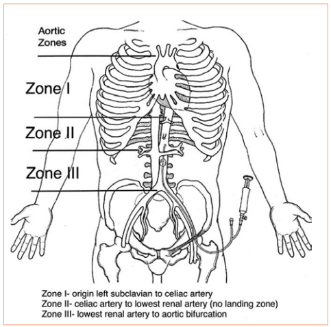
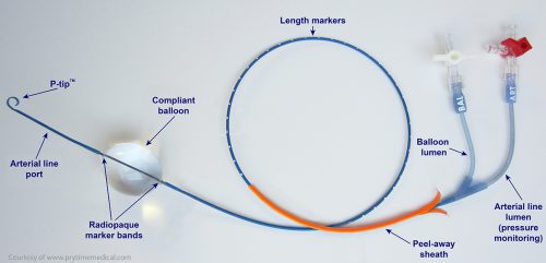
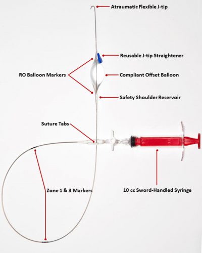
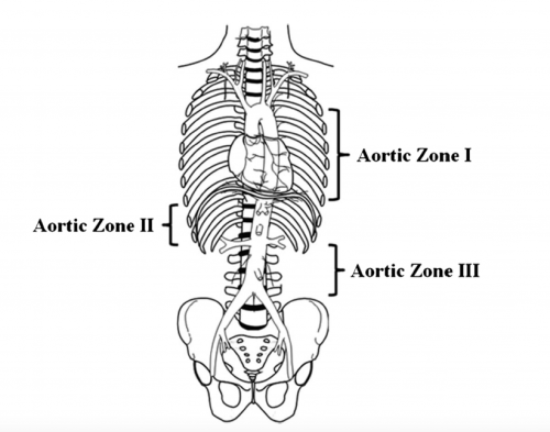
Papers for Resuscitative Endovascular Balloon Occlusion of the Aorta (REBOA) use in trauma patients have been accumulating for the past decade or so. There are three zones within the aorta, and the REBOA balloon can be inflated in either Zone II for abdominal vascular injuries or Zone III for pelvic bleeding. Here’s a nice diagram for reference:

The original studies focused on Zone II deployments, but over the past five years or so there has been growing focus on using REBOA in Zone III. Pelvic arterial bleeding can be quite problematic, and if the patient is hypotensive in the emergency department, it is not permitted to take them to interventional radiology for management. The only choice is a trip to OR for preperitoneal packing or some less effective procedure.  With the use of Zone III REBOA, it became possible to stabilize vital signs and then allow transport to IR or hybrid OR for angioembolization.

After the initial rush of very positive research, more recent papers are a bit more tempered with results that are not as cut and dried. This abstract from Hartford Hospital attempts to add a bit more information about the use of Zone III REBOA in patients who required a hemorrhage control procedure. The authors performed a retrospective review of four years of TQIP data. They compared outcome data in hypotensive adult patients with pelvic fractures requiring some type hemorrhage control procedure. The authors divided patients into REBOA+ and REBOA- groups, and looked at mortality, blood utilization, lengths of stay, and REBOA complications.

Here are the factoids:

  • Of the 4,453 patients who met inclusion criteria, only 139 patients underwent Zone III REBOA
  • The REBOA+ patients had lower BP and GCS and ISS was higher prompting the use of propensity matching
  • In order to equalize comparisons patients were propensity matched the the variables listed above, yielding 121 pairs for study
  • In-hospital and 24-hour mortality were double in the REBOA+ group (50.5% vs 25% and 31% vs 14.3%, respectively)
  • Blood transfusion was also higher in the REBOA+ group (median 4L vs 1.75L)
  • AKI was higher in the REBOA+ group (16% vs 7%) but the amputation rates were the same (no numbers given)

The authors concluded that Zone III REBOA appears to have worse outcomes and suggest that more prospective studies are indicated.

Bottom line: I have been a REBOA skeptic for some time so I have to be careful not to feed my confirmation bias. Many of the previously published positive papers include authors who have a relationship with one of the major REBOA device manufacturers. Papers from centers without any conflicts of interest are generally less positive.

Despite my own bias I also have some major questions about this abstract. The biggest thing is that I can’t make the statistics work. Granted, the entire analysis is not in the abstract. But the mortality rates given in percentages don’t yield integers when multiplied by the 121 patients in each group.

This makes me worry that we are not seeing all the statistics and we are somehow getting relative risk rather than absolute risk. I’m also confused about why the SBP, ISS, and GCS matched groups would have a difference in AKI rates in addition to the mortality numbers. Are there other significant variables affecting morbidity and mortality that were not identified or controlled?

Here are my comments and questions for the authors / presenter:

  1. Review the mortality calculations for us. How can you have a hospital mortality rate of 50.5% with 121 patients (= 61.105 people)? Provide the absolute mortality numbers so we can do the math.
  2. Why are the transfusion numbers so much higher in the REBOA+ group? Isn’t this device supposed to reduce bleeding? It seems unlikely that REBOA is making them bleed more. Is there something else going on?
  3. Similarly, why would AKI be higher in REBOA+ patients? The balloon is located below the kidneys and they should benefit from better perfusion.
  4. Could there be other factors not analyzed that contributed to the poorer outcomes in the REBOA+ group? What might they be?

I suspect the snapshot that the TQIP data allows may not be enough to let us see the entire picture here. I am looking forward to additional information during the presentation to help clarify these issues.

Reference: DOES THE USE OF REBOA IMPROVE SURVIVAL IN PATIENTS WITH PELVIC FRACTURES REQUIRING HEMORRHAGE CONTROL INTERVENTION? AAST Plenary paper #46, AAST 2022.

First, There Was REBOA, And Now… GROA?!

REBOA (Resuscitative Endovascular Balloon Occlusion of the Aorta) is a “newer” resuscitative technique that has actually been around since the Korean War. It was first used to treat two injured soldiers, and although they ultimately died, it spurred research into the technique and its applications.

Balloon occlusion techniques were then adopted by vascular surgeons and were found to be useful as an adjunct in managing ruptured abdominal aneurysms. A slow trickle of studies on human use in trauma began to surface. But when autopsy studies carried out during the Gulf War showed that uncontrolled torso hemorrhage was a major cause of death, research in the technique exploded.

First, there were a rash of pig studies evaluating the feasibility of using a percutaneously placed occlusion device in the early 2010s. This transitioned to human studies around 2014, and after that we were off to the races. Over 100 papers on REBOA are now published each year.

REBOA has been shown to have some advantage in patients with abdominal or pelvic sources of bleeding. The catheter is inserted in the groin and the balloon inflated in one of two zones, depending on the location of the hemorrhage (see diagram below). For abdominal bleeding, it is inserted just above the diaphragm in Zone I. For pelvic bleeding, it is inserted below the takeoff of the visceral arteries and above the aortic bifurcation, in Zone III.

In the US, REBOA catheters are only inserted upon arrival to the hospital. There are a few random reports of field placement where a physician is part of the prehospital team. By definition, this technique is generally not available in austere environments, only upon arrival in the emergency department.

Researchers at the University of Michigan began looking for an alternative technique that could be applied in the field by non-physicians. They noted the close anatomic relationship of the distal esophagus, proximal stomach, aorta, and thoracic vertebrae, and designed a device to compress the aorta against the spine in this area.

They developed a prototype device which they named GROA (gastro-esophageal resuscitative occlusion of the aorta). It consists of a gastro-esophageal tube with an ovoid balloon, an air pump with pressure measurement device, and an external compression device. Here is a picture of the device:

And here’s a diagram of what it looks like when inserted:

The tube is inserted and the balloon inflated. The external compression device is then placed around the patient, with the top plate located over the epigastrium and the bottom plate under the patient. It is designed to apply anterior pressure over the balloon, but to avoid circumferential constriction of the abdomen.

Bottom line: This device is an interesting development in the balloon occlusion space. As with early studies of REBOA, GROA is currently being investigated using a pig model. If it appears to be beneficial, it will still be several years before it makes the jump to human subjects. If effective, this concept would allow prehospital providers to apply some degree of hemorrhage control when it originates in the abdominal cavity.

There are currently exactly three papers on this new technique, and I have included the references below if you are interested in reading them. I’m sure there are many more to come and it may eventually be competing with REBOA for journal space.

There is one consideration to be aware of when reading these papers that is similar to much of the research on REBOA. Two of the authors have a financial interest in the company that licenses the GROA technology. And in the most recent study, another one of the authors is an advisory board member for one of the manufacturers of REBOA catheters. For these reasons, it is less likely that they will publish papers that are not favorable to the product. So read critically!

References:

  • Gastroesophageal resuscitative occlusion of the aorta: Physiologic tolerance in a swine model of hemorrhagic shock, Journal of Trauma and Acute Care Surgery: December 2020 – Volume 89 – Issue 6 – p 1114-1123 doi: 10.1097/TA.0000000000002867
  • Gastroesophageal resuscitative occlusion of the aorta prolongs survival in a lethal liver laceration model, Journal of Trauma and Acute Care Surgery: May 2022 – Volume 92 – Issue 5 – p 880-889 doi: 10.1097/TA.0000000000003444
  • Tandem use of Gastroesophageal Resuscitative Occlusion of the Aorta followed by REBOA in a Lethal Liver Laceration Model, Journal of Trauma and Acute Care Surgery: June 10, 2022 –  doi: 10.1097/TA.0000000000003719

REBOA For Pelvic Fractures

Resuscitative Endovascular Balloon Occlusion of the Aorta (REBOA) is one of the newer shiny toys that trauma professionals have adopted over the past 8 years or so. It is used to buy time for a patient who is near arrest in order to temporarily stop bleeding and get them to the operating room.

And as with all new toys, everyone wants one! I have always advised caution. Adopt a data-based approach to toy usage. Unfortunately information has been accumulating ever so slowly on this one. To help remedy this, the AAST created a registry in 2013 to consolidate the low REBOA experience numbers accruing across the US.

A group of seven surgeons representing higher-volume REBOA centers collaborated to review the AAST AORTA registry, which prospectively collects data on patients who undergo aortic occlusion. They retrospectively reviewed over six years of data on adult patients receiving REBOA for pelvic injury. They examined demographic, procedural, and outcome data in patients who underwent this procedure, both with and without other interventions like preperitoneal packing, angioembolization, or external fixation. For inclusion in the study, patients needed to have sustained blunt trauma and survived beyond the emergency department.

Here are the factoids:

  • Of the 207 patients with pelvic (Zone 3) REBOA in the registry, only 160 met inclusion criteria
  • Patients who only had REBOA suffered a mortality rate of 40% (5% in OR and 35% in the ICU)
  • Patients who had REBOA plus one of the other interventions had a 31% mortality rate (6% in OR and 25% in ICU)
  • Patients who had REBOA plus two other interventions also had a 31% mortality rate (12% in OR and 30% in the ICU)
  • Adding external fixation with or without another adjunct appeared to decrease mortality by half (from 50% to about 25%)
  • Complications were very common in all subsets, ranging from 35% to 86%
  • Patients receiving more interventions typically were more severely injured
  • No combination of REBOA and adjuncts was superior, but addition of an external fixator did appear to improve survival
  • Patients receiving angiographic embolization had a higher incidence of AKI, sometimes resulting in the need for dialysis
  • There were no significant outcome differences with REBOA use alone or with additional adjuncts

Bottom line: This was a primarily a descriptive study of how REBOA is integrated into pelvic fracture care at select US trauma centers. It was not really designed to compare the efficacy of REBOA vs preperitoneal packing vs angioembolization vs external fixation of the pelvis.

But it does show that survival remains dismal in these patients and the complication rates of REBOA + adjunct use are considerable. The authors correctly conclude that REBOA is being used in the treatment of pelvic fractures, frequently with the addition of other adjuncts. They state that the benefit of more interventions must be balanced against the potential for complications. And finally, they note that there is a need to fill in the evidence base if we are ever to adopt REBOA as a standard of care for select pelvic fractures.

What does this mean to all of you who are thinking of playing with this toy? Proceed with caution! The learning curve is steep. The complication rate is high. The opportunity for mayhem is great. This means that you must proceed deliberately. Get some advanced training with this technique. Use your performance improvement program to impartially critique its use with every deployment. And submit your experience to the national registry so we can all learn from your experience and figure out how to optimize use of this tool.

Reference: Patterns and outcomes of zone 3 REBOA use in the management of severe pelvic fractures: Results from the AAST Aortic Occlusion for Resuscitation in Trauma and Acute Care Surgery database. J Trauma 90(4):659-665, 2021.


REBOA: A Comparison Of The Hardware From Two Companies

I started off the week describing a study using a new version of the REBOA catheter (Resuscitative Endovascular Balloon Occlusion of the Aorta) that was smaller than the more commonly used one. Today I’ll put both side by side and describe the similarities and differences.

First, let’s start with the current market leader, the ER-REBOA catheter by Prytime Medical in Boerne, TX. Here’s a picture provided by the company:

And here’s a photo of the Frontline Medical Technologies COBRA-OS, based in London, Ontario, Canada. This acronym stands for Control of Bleeding, Resuscitation, Arterial Occlusion System. Now, REBOA is used by surgeons as a general descriptor for this type of technology. I assume that Frontline does not include REBOA in the name of this product since Prytime has incorporated it into theirs.

There are a number of similarities, as well as some key differences. Let’s start at the tip and make our way back to the syringe.

Catheter tip: Prytime has a trademarked “P-tip” which has a little extra curl compared to the Frontline’s flexible j-tip. The Prytime version is designed to “help reduce catheter migration and aid in positioning. Although a guidewire can be inserted into either to assist in repositioning, it does not enter the P-tip. And note, neither device requires a wire for insertion.

Arterial line port: This is only found on the Prytime device. This is located just distal to the balloon so arterial pressures can be measured above the catheter after inflation. This port extends through the catheter, terminating in a hub that can be connected to standard pressure transducer equipment. The Frontline device is too small to incorporate this feature.

Balloon: The Prytime balloon is a more standard ovoid shape. The company provides guidelines of 8cc inflation for Zone I and 2cc for Zone III. This can be adjusted based on confirmation of occlusion provided by the arterial pressure wave form. The Frontline device has an “ice cream cone shaped” balloon with the taper proximally and a “safety shoulder” to protect the balloon. The company claims that this design helps reduce the likelihood of rupture. The balloon will accept 13cc at maximum inflation. Since there is no arterial line, alternate means (palpation, ultrasound, or a transducer in the insertion port) must be used to determine degree of occlusion.

Markers: The Prytime device has radio-opaque markers at either end of the balloon, as well as length markers on the proximal portion of the catheter. The Frontline catheter has the same markers around the balloon, but only two large visible marks on the proximal catheter. These are marked for placement in Zone I (48cm) and Zone III (28cm) in average size patients.

Sheath: The Prytime product has a peel-away sheath that is used to cover the P-tip to straighten it. This unit is then inserted into the previously placed access port. Once inserted the sheath is peeled away after the balloon has passed the end of the port. The Frontline device does not have a sheath, but includes a reusable j-tip straightener on the catheter. This straightens the tip as it passes through the port.

Access port: These are included with both products and are inserted using typical Seldinger technique. Both have a side port for fluid infusion. The side port of the Frontline product can be used as an arterial pressure monitor. The port is 7Fr in the Prytime product and 4Fr for Frontline. This smaller size may decrease the incidence of vascular thrombosis or vessel injury requiring repair after removal.

Bottom line: I’ve described two different products that allow trauma professionals to use the REBOA concept. This evolution demonstrates the usual cycle of new product and feature refinement that we have come to expect in medical devices.

Is one “better” than the other? That’s probably not the right question. More likely, it will boil down to which one is right for a particular patient or situation. Only time, and lots of additional research, will tell.

References: 

  • Prytime Medical – www.prytimemedical.com
  • Frontline Medical Technologies, Inc. – www.frontlinemedtech.com

I have no financial interest in either of these companies

The Shrinking REBOA Catheter

REBOA (resuscitative endovascular balloon occlusion of the aorta) is one of the relatively new toys in our trauma toy chest. Although it’s been used for decades by vascular surgeons, believe it or not it only made the jump into the trauma world less than 10 years ago.

The original catheters used during the early days, and primarily in swine models, required a 15 French sheath for insertion. As might be expected, insertion of these huge sheaths into the common femoral artery can cause significant vascular injury. Equipment manufacturers have been steadily reducing the size of the REBOA catheter, first to 12 Fr and then to the 7 Fr size commonly used today.

The surgery group at the London Health Sciences Center in London, Ontario, Canada performed a pilot study of a new and much smaller sheath and REBOA catheter. It is made by Front Line Medical Technologies, also located in London. This was a proof of concept for the device and was performed in seven neurological death organ donors prior to their donation.

The kit consists of a 4 Fr sheath introducer with a 21 gauge needle and a guidewire, plus the REBOA catheter itself. Here is an image of the catheter:

This catheter includes several innovations not found in current catheters used in the US. I will do a side by side review of these later this week.

Here are the factoids:

  • Seven organ donors were studied after appropriate consent from the hospital IRB, organ procurement agency, organ donor procurement team, and family
  • A single general/vascular surgeon performed all insertions
  • A left sided arterial line using the 4 Fr sheath was inserted for monitoring before the procurement began
  • A right sided 4 Fr sheath was inserted for catheter insertion after the procurement incision was made
  • Average sheath insertion time was 48 seconds, and deployment time for the catheter was an average of 70 seconds (max time was 105 seconds)
  • Occlusion was confirmed by the left femoral arterial pressure monitor and by palpating the aorta below the baloon

Bottom line: This was a very simple study of the feasibility of using a smaller REBOA catheter. It measured both ease of insertion and presence of full occlusion. This is an exciting study, because there is the potential for easier insertion and fewer vascular complications at the insertion site. Obviously, these factors are not yet known, and only further work will make this clear. 

Nonetheless, easier and safer insertion has the potential to increase the use of REBOA. This will allow us to get quicker answers to the nagging questions about whether it is actually a valuable resuscitation tool and help us figure out how and in whom it is best used.

Reference: Size matters: first-in-human study of a novel 4 French REBOA device. Trauma Surgery & Acute Care Open 2021;6:e000617. doi: 10.1136/tsaco-2020-000617