Tag Archives: TBI

Best Of EAST 2024 #9: The Burden Of Transferred TBI Patients

In theory, tiered trauma centers should allow patients with lesser injuries to be treated at lower levels and more severe trauma at higher-level centers. This parallels the resource availability at those centers. In reality, many patients with injuries that seem complex (solid organ, children, and TBI) are transferred due to a “lack of comfort” in taking care of them or the perception that they may deteriorate quickly.

The truth is that many, if not most, of these patients are discharged shortly after transfer to the higher-level center, with minimal intervention. This burdens the trauma system in several ways. First, trauma professionals at the lower-level centers slowly lose their skills and comfort in taking care of these patients. The prehospital system is already plagued with low resources and a shortage of personnel. Using one of the only ambulances in a rural area for transport to a distant center takes it out of the community, potentially putting the area population at risk for delayed care.

The University of Arizona at Tucson group performed another TQIP study to highlight this problem. They performed a four-year retrospective analysis of transfer data in patients with isolated TBI with intracranial hemorrhage. They observed the number of transferred patients who required CT scans, ICP monitoring or craniotomy/craniectomy, length of stay, and mortality.

Here are the factoids:

  • Of the nearly 120,000 patients with isolated TBI at Level I and II centers, 45% were transferred from other centers
  • Most patients had GCS 14-15 on arrival, but 10% had GCS 8
  • CT was performed in 58%, and another repeat CT in 4%
  • Four percent underwent ICP monitoring, and 12.5% had a crani
  • Mortality was 6.5%
  • Median length of stay was two days, with a range of 1-5
  • 18% were discharged within 24 hours, and 39% within 48 hours

The authors concluded that, while half of isolated TBI patients were treated at high-level trauma centers, one-third were discharged home within 48 hours with no intervention other than a CT scan. They recommend systemwide guidelines to improve resource utilization.

Bottom line: This straightforward analysis highlights one of the most significant issues facing trauma systems: unnecessary transfer. For decades, Level I and II centers were convenient and always available for transfer, even if the indications were questionable. Then COVID came along and changed everything.

Now, resources are tight everywhere. EMS is underpaid and under-reimbursed, and personnel are difficult to recruit. Hospital personnel of all types face low staffing levels, making it more stressful to provide the level of care we are accustomed to. Skilled nursing facilities, rehab centers, and other outpatient care settings face the same problems.

This has created a domino effect, where the lower-level centers want to transfer a patient but can’t find a bed at the higher-level centers. When they do, it takes forever to get them transported. Then, the higher-level centers can’t discharge them if they need any level of care other than home care.

There are many pieces to this puzzle, but this abstract clearly outlines one of them. Lower-level centers are transferring some patients who could actually be admitted to them. Several reasons may be given, but it typically boils down to the surgeons or the hospitalists not being “comfortable” with certain patients or worrying that they could deteriorate.

This paper does not tease out what kind of isolated TBI the patients had, and I recommend they do. There is a big difference in patients with a subarachnoid hemorrhage (SAH) vs. those with an epidural. The number of patients with SAH is far greater, and the vast majority can go home after a brief (or no) observation. The likelihood of deterioration in patients not on blood thinners is nearly zero. 

State trauma systems and higher-level trauma centers should work with their Level III and IV partners to adopt consistent practice guidelines and protocols to stratify these patients to identify those at low risk. The higher-level centers should provide education to help their referral partners develop a baseline comfort level with these patients. This is the only way we can begin to realign the levels of trauma centers with the levels of care needed by our patients.

Reference: Endless highways: the burden of transferred traumatic brain injury patients in the United States. EAST 2024, Podium paper #42.

Best Of EAST 2023 #12: VTE Prophylaxis In Severe TBI

Time for another abstract on venous thromboembolic disease (VTE) prophylaxis, but this time in patients with severe head injury. VTE is a significant problem for trauma patients. Those with a potential source of bleeding from their injuries cause us to hesitate and consider the timing of chemical prophylaxis closely. Do we really want to cause more bleeding?

This is particularly problematic with intracranial hemorrhage, as the treatment is major brain surgery. Over recent years, the literature has been leaning toward earlier prophylaxis as soon as the intracranial blood has stopped evolving.

The EAST Multicenter Trials Group performed a seven-year retrospective review at 24 Level I and II trauma centers to assess the safety and efficacy of VTE chemoprophylaxis.  They divided patients into three groups: no prophylaxis, early prophylaxis (within 24 hours), and late prophylaxis (after 24 hours).

The authors assessed two endpoints: VTE occurrence and expansion of intracranial hemorrhage (ICH). They used several regression models to check their hypotheses.

Here are the factoids:

  • A total of 2,659 patients met the inclusion criteria. This averages out to 15 eligible patients per month per center. This is probably reasonable when combining a few high-volume centers with more lower volume centers.
  • Compared to early prophylaxis, patients who received late prophylaxis were twice as likely to develop VTE, although this was not statistically significant (p = 0.059)
  • Compared to early prophylaxis, patients who received no prophylaxis were a third less likely to develop VTE, although this, too, was not statistically significant (p = 0.39
  • About 25% of patients who received either early or late prophylaxis suffered an extension of their ICH, but only 17% of the no-prophylaxis group did
  • The regression model showed that the no prophylaxis group was 36% less likely to develop ICH extension compared to either early or late prophylaxis groups.

The workgroup concluded that the development of VTE was not dependent on the timing of the start of prophylaxis. Furthermore, patients who did not receive any prophylaxis had significantly decreased odds of ICH extension. The group recommended larger randomized studies to extend this work.

Bottom line: Shocker! This multicenter study suggests that the no prophylaxis and early prophylaxis groups had fewer VTE events than the late group, although these results were not statistically significant. This means that there wasn’t an advantage to giving the shot.

And the other major conclusion was that both early and late prophylaxis was associated with a significantly higher incidence of ICH extension. 

Roll these together, and you will find that neither early nor late prophylaxis help prevent VTE, yet they are both associated with additional bleeding in and around the brain! 

Heresy! I am trying to figure out what to make of these results. Perhaps the retrospective nature of the study and the wildcards this introduces influenced the results. It could be a study power problem, except the numbers were approaching significance that was unfavorable for prophylaxis.

I will be very interested to hear how the authors explain these findings. And yes, a well-powered randomized study would be great, but I don’t think many institutional review boards will be keen on a no-treatment group given our current fear of VTE. So don’t count on any real answers soon.

Reference: EARLY VTE PROPHYLAXIS IN SEVERE TRAUMATIC BRAIN INJURY: A PROPENSITY SCORE WEIGHTED EAST MULTICENTER TRIAL. EAST 2023 Podium paper #38.

Best Of EAST 2023 #4: Whole Blood In Patients With Shock And TBI

We know that even a brief shock episode in patients with severe TBI dramatically increases mortality. Therefore, standard practice is to ensure good oxygenation with supplemental O2 and an adequate airway ASAP and to guard against hypotension with crystalloids and blood if needed.

Many papers (and several abstracts in this bunch) have been written about the benefits of whole blood transfusion. The group at the University of Texas in Houston compiled a prospective database of their experience with emergency release blood product usage in patients with hemorrhagic shock.

They massaged this database, analyzing a subset of patients with severe TBI, defined as AIS Head of 3. They specifically looked at mortality and outcome  differences between those who received whole blood and those who received component therapy.

Here are the factoids:

  • A total of 564 patients met the TBI + shock criteria, and 341 (60%) received whole blood
  • Patients receiving whole blood  had higher ISS (34 vs. 29), lower blood pressure (104 vs. 118), and higher lactate (4.3 vs. 3.6), all indicators of more severe injury
  • Initial univariate analysis did not identify any mortality difference, but using a weighted multivariate model teased out decreases in overall mortality, death from the TBI, and blood product usage
  • Neither statistical model demonstrated any difference in discharge disposition of ventilator days

The authors concluded that whole blood transfusion in patients with both hemorrhagic shock and TBI was associated with decreased mortality and blood product utilization.

Bottom line: This is yet another study trying to tease out the benefits of giving whole blood. The results are intriguing and show an association between whole blood use and survival. But remember, this type of study does not establish causality. It’s not possible to rule out other variables that were not available or not considered that could be the cause of the difference.

In this type of study, it’s essential to look at the design. Was it possible to create the study to record a complete set of variables that the researchers thought might contribute to the outcomes? Or is it a retrospective analysis of someone else’s data that contains just a few of them? This study falls into the latter category, so we have fewer data elements to work with and the likelihood that others that are not present could contribute to the outcomes.

The details of the multivariate analysis are also important. The authors stated that weighted multivariate analyses were performed. It’s not possible to provide details in a standard abstract, but these will be important for the audience to understand.

Here are my questions and comments for the presenter/authors:

  • Tell us more about the database you used for the analysis. What was the purpose? How many data elements did you collect, and how are they related to your research questions?
  • How did you decide which variables to include in your multivariate analysis? And how did you determine the weights? These can have a significant effect on your results.
  • This is a preliminary proof of idea study. How should this be followed up to move from association to causation?

This is just one of many exciting studies trying to shed light on the forgotten benefits of whole blood in trauma. I’m looking forward to seeing the final manuscript!

Reference:  PATIENTS WITH BOTH TRAUMATIC BRAIN INJURY AND HEMORRHAGIC SHOCK BENEFIT FROM RESUSCITATION WITH WHOLE BLOOD. EAST 2023 Podium paper #2.

What GCS Should Trigger Trauma Activation?

For the most part, trauma centers are free to pick and choose their own trauma team activation trigger criteria. Typically, these are a mix of physiologic, anatomic, and mechanistic items. However, the American College of Surgeons Committee on Trauma mandates that either seven (Orange Book) or eight (Gray Book) specific criteria must present in every center’s highest-level activation list.

One of these mandatory criteria is a Glasgow Coma Scale (GCS) score of eight or less. The reason is that this level denotes a severe brain injury and as patients approach it they are less and less able to protect their own airway. Although this specific GCS is a minimum, centers are free to choose their own specific threshold as long as it is not any lower.

How does a center choose the “right” GCS? It seems straightforward, right? A mild TBI is defined as GCS from 13-15. These patients have only lost one or two points in their eye-opening, verbal, and motor scores and are relatively unlikely to have a significant lesion in their head or an airway issue.

At the other end of the spectrum is the severe TBI, with a GCS of 3-8. These are a chip shot, with the potential for severe injury and a frequently threatened airway. They demand rapid assessment and intervention, hence the required trauma activation.

But what about those patients with moderate TBI with a GCS from 9-12? They obviously have a higher risk for serious intracranial injury. And as the GCS declines, the patient’s ability to protect their airway decreases. At some point between those GCS scores, most clinicians hit their own internal trigger to provide a definitive airway.

So what do actual trauma centers choose as their threshold? I conducted an informal survey of my readers, asking them to provide their specific GCS threshold.

Here are the factoids:

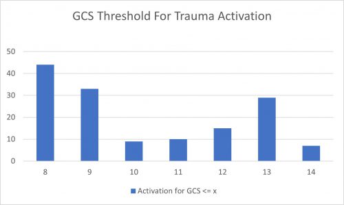
  • A total of 147 trauma centers of all levels responded
  • They were located in the United States, Germany, Saudi Arabia, and Singapore
  • This chart shows the number of centers that selected a threshold less than or equal to the GCS on the horizontal axis:


  • Nearly a third of centers (30%) adhere strictly to the ACS criterion of 8
  • Another 22% use a threshold of 9, possibly to avoid any confusion from having a “less than or equal to” criterion
  • There is another bump on the curve at 13, with 20% using this threshold

Bottom line: A little more than half of centers use a GCS threshold of 8 or 9 as their TTA trigger. This meets the ACS criteria, but could potentially leave a few airways unprotected from time to time. Only about 5% of centers use the higher GCS levels with the exception of GCS 13. That seems to be another popular one.

Which one is right for you? GCS 8 will always work because it is the minimum requirement. My own personal threshold trends higher. I would rather be called to an activation and apply my own judgement rather than come running only when the patient needs to be intubated followed by a trip to the OR for craniotomy.

You will need to work with your emergency physicians, trauma surgeons, and neurosurgeons to determine their collective comfort levels. It comes down to a balance between safety and unnecessary intubation. Look at your own center’s experience and pick a threshold that achieves a proper balance of overall patient safety.

Stroke And Fall VS Fall And Bleed

It’s like the old chicken and egg question. When dealing with head trauma and falls, which came first? Did the patient have a stroke and then fall down? Or did they fall and sustain some type of intracranial hemorrhage? And you may ask, does it make a difference? They are going to get a head scan anyway, right?

In my opinion, it makes a big difference! How often have you seen the following scenario? EMS is called to a house or nursing home for someone who has fallen. They notice some extremity weakness on one side and presume the patient is having a stroke. The emergency department is then notified that a stroke patient is inbound.

On arrival, the patient was rapidly assessed and whisked off to CT scan for a CT and angiogram, possibly with neurology present. My experience is that a majority of these scans is negative for CVA. And many are positive for some type of extra-axial hemorrhage like subdural or epidural blood from the real injury.

Unfortunately, something called anchoring bias is likely to occur in this situation. Everyone from the paramedics onward are moving along under the assumption that the patient has had a stroke. They stop considering the more common diagnosis of TBI and other potential injuries in the spine and torso. Even when the CT angiogram is found to be negative, it’s difficult for people to change gears. It then takes longer to address the subdural or epidural. The involved trauma professionals are less likely to activate the trauma team. And further evaluation of the chest, abdomen, and spine may be delayed or forgotten for a time.

Bottom line: In any case of a fall followed by neurological changes that could indicate stroke, always presume a serious TBI first! If EMS requests a stroke code, it should be changed to a trauma activation prior to patient arrival. This takes advantage of the odds (more in favor of TBI) and activates a team that is well versed in evaluating the entire patient. If no evidence of hemorrhagic stroke is present, the team will then order the brain CTA and involve the stroke team as necessary.

And for good measure, every one of these cases that does start as a stroke evaluation should be addressed by the trauma performance improvement process!