Category Archives: Imaging

How To Detect Bucket-handle Intestinal Injuries With CT Scan

A bucket-handle injury is a relatively uncommon complication of blunt trauma to the abdomen. It only occurs in a few percent of patients, but is much more likely if they have a seat belt sign.  The basic pathology is that the bowel mesentery (small bowel of sigmoid colon) gets pulled away from the intestinal wall.

This injury is problematic because it may take a few days for the bowel itself to die and perforate. Patients with no other injuries could potentially be discharged from the hospital before they become overtly symptomatic, leading to delayed treatment.

Here’s an image from my personal collection with not one, but four bucket-handle injuries.
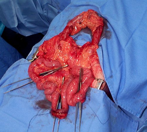
Typical patients with suspected blunt intestinal injury are observed with good serial exams and a daily WBC count. If this begins to rise after 24 hours, there is a reasonable chance that this injury is present.

CT scan has not really been that reliable in past studies. There may be some “dirty mesentery”, which is contused and has a hematoma within it. But without a more convincing exam, it is difficult to convince yourself to operate immediately on these patients.

A paper was published by a group of radiologists at Duke University. It appears to be a case report disguised as a descriptive paper. It looks like they picked a few known bucket-handle injuries from their institution and back-correlated them with CT findings.

The authors called out the usual culprits:

  • Fluid between loops of bowel
  • Active bleeding in the mesentery
  • Bowel wall perfusion defects

But they also noted that traumatic abdominal wall hernias were highly with the injury as well. These are rare, but should bring intestinal injury to mind when seen.

With newer scanners, radiologists are better able to detect subtle areas of hypoperfusion as well. This is a fairly good indicator of injury, especially when adjacent bowel appears normally perfused. Here are two examples. The black arrows denote active extravasation, and the white ones an area of hypoperfusion.
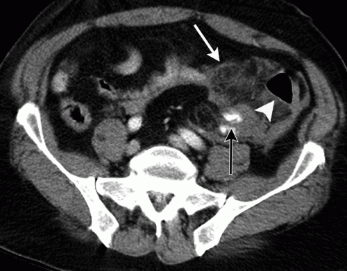
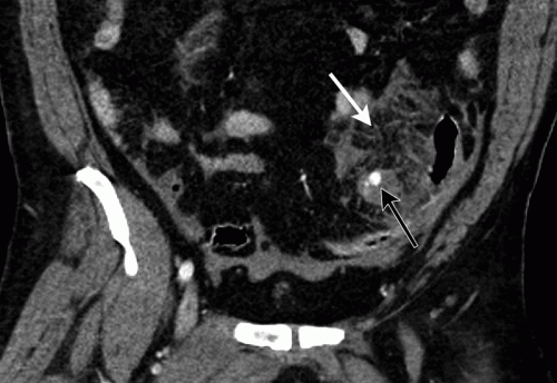
The authors add bowel wall hypoperfusion as another finding that may point to a bucket-handle type injury

Bottom line: Hold the phone! Don’t change your practice yet. This paper is not able to demonstrate how good this radiographic sign is. Looking at other radiology literature, the specificity is about 90%. But remember, that means that if they don’t have the CT finding, that’s true only 90% of the time.

Unfortunately the sensitivity is only 10%. So if you see it on the scan, they’ve got a 1 in 10 chance of actually having the injury. That’s not good enough for me to run to the operating room.

Here’s what I recommend: if your patient has an unconcerning exam and any of the usual culprits (pelvic fluid, inter-loop fluid, dirty mesentery, thickened bowel loops, abdominal wall hernia), perform serial exams and get a WBC the next morning. If the exam worsens, operate. If the WBC rises, consider laparoscopy to see if you need to make a bigger incision. And if you see this new kid on the block, the hypoperfused bowel, consider laparoscopy right away. 

I’m sure the radiologists and the technology will keep getting better. But for now, blunt intestinal injury still requires patience, perceptiveness, and a little luck.

References:

  • CT findings of traumatic bucket-handle mesenteric injuries. Am J Radiol 209:W360-@364, 2017.
  • Multidetector CT of blunt abdominal trauma. Radiology 265(3):678–693, 2012.

Is The Trauma Bay Chest X-Ray Really Necessary Or Just Dogma?

I love challenging dogma. I spoke last week (virtually) at an excellent event at the Intermountain Medical Center in Utah. One of my talks there addressed trauma myths and dogma.

I bring this up because there is an interesting article in the Journal of Trauma this month that questions the necessity of the routine chest x-ray (CXR) in blunt trauma resuscitation. So of course, this caught my eye. Let’s dig in.

The first thing to understand is that this article is an opinion piece and is identified as such. It was written by three surgeons, including the trauma medical director, at the Stanford University Hospital trauma center.

First, what are we really looking for on the chest x-ray that is taken in the trauma bay? I call them “the three big things”.

  • Big air. The first item to be identified is a pneumothorax. The chest x-ray helps the trauma professionals decide if the pneumo needs an intervention (chest tube) and when. (Note: it could in theory identify a tension pneumothorax. But in that case, the trauma pros should be embarrassed. They should have picked that up on their clinical exam and assessment of the vitals.)
  • Big blood. The chest x-ray can also identify a hemothorax. And once again, it can help decide whether its size warrants chest tube insertion.
  • Big mediastinum. A wide mediastinum may indicate the presence of hematoma from an aortic injury. It is one of the indications for performing CT angiography of the chest to rule it out.

Here are their authors’ arguments:

  • There are other imaging modalities available to us that are very accurate. FAST ultrasound has been used routinely for abdominal and cardiac evaluation for over a decade. The extended FAST (eFAST) involves evaluation of the pleural interface to identify pneumothorax. A study published last year pitted CXR vs eFAST. It found that the eFAST outperformed with a sensitivity of 94% and specificity of nearly 100%.
    But what about hemothorax? Ultrasound is less helpful here. But the CT scanner is. It is far more accurate at identifying and quantifying hemothorax than the CXR.
  • Evaluation of the aorta can either wait, or it can’t wait at all. If the patient loses vital signs in the trauma bay the decision to open the chest or insert a REBOA catheter must be made. In the latter case, a chest x-ray must be obtained to exclude a thoracic source of bleeding that the cathether is of no use for. But if the patient truly is bleeding out from a blunt aortic injury, it is nearly certain that he or she is not leaving the trauma bay alive.
    What about using the wide mediastinum as an indication or order the chest CT angiogram? The authors argue that there will probably be a history of deceleration or other associated injuries (femur fracture is a very common one).

Bottom line: The authors argue that the chest x-ray should go the way of the lateral cervical spine x-ray used at the turn of the 21st century and before. They claim that judicious use of the extended FAST and CT angiography can identify the significant injuries we need to know about in a timely manner.

My own opinion is more nuanced. I buy their arguments that the extended FAST will identify all significant pneumothoraces. However, we have typically answered the question “how big is too big” using the chest x-ray. That is the most helpful tool in deciding whether a chest tube is warranted or not.

As for hemothorax, I don’t believe that a CT is the best tool for evaluating this, either. Are the authors members of the “pan-scan” school? What about those of us that use the “selective scan” philosophy. True, the abdominal scan will identify both hemothorax and pneumthorax on the lower cuts of the chest. But as in the previous paragraph, we are better trained to judge when a chest tube is indicated by the appearance of the chest x-ray. Hemothorax (or pneumothorax) is not an indication to get a chest CT.

I don’t buy argument that there will be other indications of potential aortic injury. Deceleration is in the eye of the beholder. How do we know how fast the vehicle was actually moving? What is the magic velocity that will break this patient’s aorta? This particular patient may not have any of the other potential indicators that increase suspicion for aortic injury. That wide mediastinum may be the only clue. Yes, the numbers of affected patients are small, but the consequences of missing one could be deadly.

And what about patients who might not get scanned at all? And those who need a study to confirm tube or line placement? They must absolutely get a chest x-ray before they leave the trauma bay.

At this point, I can’t see a way to dispense with the chest x-ray completely. It should still be used to:

  • Confirm pneumothorax from eFAST to help decide if a chest tube is needed
  • Identify potential pathology (hemothorax, wide mediastinum) in patients who don’t otherwise meet criteria for chest CT
  • Verify endotracheal tube position after intubation

What do you think? Please leave your comments or Tweets about this topic.

References:

  • Extended-FAST plus MDCT in pneumothorax diagnosis of major trauma: time to revisit ATLS imaging approach? J Ultrasound. 2019;22(4):461–469.
  • Necessity of routine chest radiograph in blunt trauma resuscitation: Time to evaluate dogma with evidence. J Trauma 2020;89(3):e69-70.

Best Of AAST #11: Hard Signs Of Vascular injury

The next abstract in this series poses a challenge to long-held dogma. More than three decades ago, examination of vascular injuries was divided into “hard signs” vs “soft signs.” Hard signs consisted of findings like pulsatile hemorrhage, expanding hematoma, absent distal pulses, thrill, or bruit. These were believed to be absolute indications to proceed directly to the operating room for exploration and repair.

But now, in this day and age of CT angiography  (CTA)and all manner of endovascular techniques and tools, things seem to be changing. There is more reliance on CTA, and a willingness to image patients with hard signs before considering an operation. But is this prudent?

The AAST established the Prospective Observational Vascular Injury Treatment (PROOVIT) database as a multi-institutional effort involving major trauma centers around the country in 2013. A group based at the Massachusetts General Hospital massaged the data to study current patterns in assessment and management of patients with penetrating extremity vascular injury. Specifically, they were interested in examining the presence of hard signs and the outcomes after initial imaging and operative management.

Here are the factoids:

  • A total of 1,910 database records were reviewed, of which 1108 (58%) presented with hard signs of injury
  • 83% of the patients with hard signs had either active hemorrhage or expanding hematoma; only 15% had ischemia
  • CTA was used in a quarter of patients with hard signs (24% hemorrhagic, 40% ischemic)
  •  Two thirds of patients with hard signs were taken to OR without imaging (70% hemorrhagic, 45% ischemic)
  • Open repair was performed in about two thirds of hemorrhagic and ischemic patients both with and without imaging, but endovascular  or hybrid repairs were 5x more likely (2% vs 10%) in patients who underwent imaging first
  • There were no differences in outcomes (amputation, mortality, blood transfusions, reoperation) between the open and endovascular/hybrid repair groups

The authors concluded that stable patients with hard signs of vascular injury may benefit from preop imaging to help plan the specific mode of repair to be performed (open vs endovascular / hybrid).

Here are my comments: This was a retrospective review of prospectively collected data. The database has a wealth of detail, and this is a simple and clean analysis of a specific question. The results and analyses were straightforward and easy to follow.

What this study does is to call into question the old dogma of rushing straight to the operating room with any patient who has hard signs of vascular injury. The advent of endovascular tools and techniques has allowed us to more easily address some vascular injuries that were previously problematic due to their location and accessibility.

Being a descriptive study only, it showed us “what we did” with vascular injuries during the time period of the database. And it also showed that the surgeons were more likely to use endovascular techniques if they were able to take the time for preop imaging. Most importantly, it demonstrated that gross outcomes like death, reoperation, and amputation were not increased by the delay needed to obtain that imaging.

I consider this to be a pilot project. And the authors correctly state that the next step is a true prospective study to confirm that this should be the new way of thinking about hard signs in the future.

Here are some questions for the presenter and authors.

  • Please provide more information on the database records used. Which years were included? What were the inclusion criteria? Were any patients excluded?
  • What was the definition of a vascular injury to the extremity? Did it include the very proximal brachial artery or the distal subclavian? These may increase the likelihood of choosing an endovascular repair.
  • Did you stratify by type of penetrating injury (stab vs gunshot) or velocity (assault rifles and shotguns)? These will increase the likelihood of proceeding directly to OR and potentially skew the data.
  • Some data from the abstract is missing, typically p values. This appears to be a glitch with the abstract entry system, since it is a problem in other abstracts as well.
  • How long do you think it will take to collect adequate data from a prospective study so that preop imaging in stable patients becomes the new standard of care?

This was a fun abstract to read! I’m looking forward to the presentation next week.

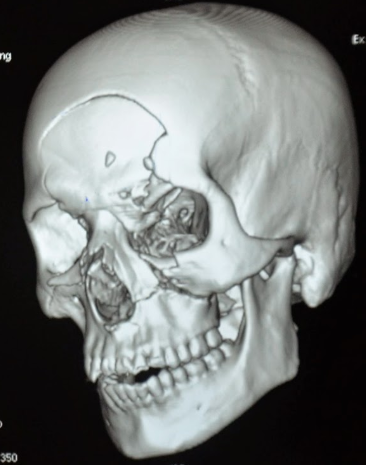
When To Obtain A Dedicated Facial CT

Initial CT scan evaluation for blunt trauma patients is fairly standardized. The usual palate consists of scans of the head, cervical spine, chest, abdomen and pelvis. Some choose their “colors” individually, and others just slop everything on the canvas.

However, there are a few other scans that are occasionally helpful and/or necessary. Think soft tissue views, or CT angiogram of the neck, or CT angiogram of potential extremity vascular injuries.

Another study that is occasionally needed but many times unnecessarily ordered is the dedicated CT of the facial bones. This study spans the entire area from mandible to frontal sinus and is performed using finer cuts to display greater detail.

The unfortunate truth is that a large number of dedicated facial CTs either do not show fractures, or show fractures that don’t require operation. The scan does deliver a nice dose of radiation, though. Is there any way to be more selective about ordering it?

About 10 years ago, a plastic surgery group in Madison developed what came to be called the “Wisconsin criteria” for ordering facial imaging.  Here they are:

  • Bony step-off
  • Periorbital ecchymosis
  • GCS < 14
  • Malocclusion
  • Missing teeth

The authors claimed 97% sensitivity and 2.6% missed fracture rate, although external validation suggested those numbers were a bit generous. The Plastic Surgery group from the University of Minnesota and Regions Hospital recently re-studied these criteria with a large number of patients, looking at accuracy as well as cost-savings.

They performed a retrospective review of 1000 patients (based on a power analysis) who had a facial CT and adequate documentation of the Wisconsin criteria in the chart. Here are the factoids in table form:

(click table for larger copy)

  • Periorbital ecchymosis was the most common criterion, which had the highest sensitivity of 70% (terrible)
  • The other criteria fared even worse from a sensitivity standpoint
  • But if you roll them all up together, the presence of any one of the five yielded a 90% sensitivity (true positives) and 52% specificity (true negatives)
  • The negative predictive value was 93% if none of the criteria were present, which means it’s a good tool for ruling out the need for a CT scan
  • The overall missed fracture rate was 2.8%, and only 0.12% for ones that required operation
  • Cost savings by limiting CT to patients who met the criteria was over $300K in 2014

Bottom line: What to do? It’s clear that using the absence of any of the Wisconsin criteria to avoid a facial CT scan is helpful. This makes sense, because 4 of the 5 criteria are findings on facial exam. But it also means that a lot of scans will still get done for low sensitivity criteria. 

How about this? Since nearly all of these patients will have head and cervical CT scans, review the head scan first for facial fractures. Single, non-displaced fractures are nearly always nonoperative in nature. If patterns of fractures are present, or there are significant displacements, a dedicated facial scan will be very helpful in determining operative management.

But remember, the head CT does not include the mandible. A good physical exam and occlusion check is mandatory, and any suspicion of injury should prompt a full scan of the face.

Thanks to Chris Stewart, the lead author on this study for sending it to me for review.

Rreference: Validation of the “Wisconsin criteria” for obtaining dedicated facial imaging and its financial impact at a Level I trauma center. Craniomaxillofacial Trauma & Recon 13(1):4-8, 2020.

What? Still Using MRI For Cervical Spine Clearance?

Cervical spine clearance as evolved considerably over the years. First, there were five views of the spine using plain radiography. Then there were three. Then we moved to CT scan with clinical clearance. And currently, many institutions are relying only on CT.

But MRI has been used as an adjunct for quite some time. Initially, it was the tie breaker in patients who had equivocal CT findings, and for a while it was used for clearance in obtunded patients. And thanks to conflicting literature and disparate studies, the occasional usage became more frequent.

The group at Cedars-Sinai Medical Center in Los Angeles  noted that the percentage of patients undergoing MRI for cervical spine evaluation at their center slowly slowly crept up from 0.9% to 5.6% over a 10 year period. They designed a study to analyze the utility of this practice and inform their future practice.

Here are the factoids:

  • Over 9,000 patients had cervical spine CT during the 10-year study period; 513 (5.6%) were positive
  • Of the 513 CT-positive patients, 290 (56%) underwent an MRI. This showed:
    • Confirmation of the major injury in 250
    • Minor injury in 40
    • Clinically significant injury was seen in only 2 which was no surprise since they both had neurologic deficits
  • Of the 8,588 CT-negative patients, only 9 had clinically significant findings and 8 of them had neurologic deficits

Bottom line: So what have we learned here? First, MRI usage at Cedars-Sinai increased over time but was really not that useful. The main use was for imaging obtunded patients or those with an obvious neurologic deficit.

More than half of patients with positive CT scans also underwent MRI. If a major injury was seen on CT, MRI confirmed it. But if the CT findings were minor, none of the MRIs added any clinically significant findings in the absence of a neurologic deficit.

And what about MRI after negative CT? In the absence of a deficit, only one had a clinically significant finding (which only required a brace).

This study shows the wisdom of monitoring “how we do it.” There is sometimes some creepage away from what the literature shows is the best practice. The best way to remedy this is to do a good study, just like the authors did. They saw a slow change in practice, investigated it, and found that there was no good clinical reason for it. This gives the trauma program the ammunition to squelch the unwelcome behavior and return the clinicians to best practices.

Reference: Is MRI becoming the new CT for cervical spine clearance? Trends in MRI utilization at a Level I trauma center. J Tra publish ahead of print, DOI: 10.1097/TA.0000000000002752, 2020.