Category Archives: Imaging

What Is: A Pars Fracture / Defect

Radiologists sure know their anatomy! The vast majority of the time, I actually know what they are describing. But every once in a while they’ll toss in some term that I know I probably learned about in medical school (last century). For whatever reason though, I’m just not able to retrieve it.

Which brings me to the pars fracture. Hmm. I figure that if I have to hit the books again to look something up, there are probably a few other trauma professionals out there who are dying to know what it is, too. Here’s a diagram of a typical vertebra:

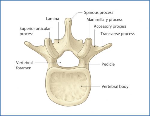
The arch extending away from the vertebral body consists of the pedicles, which are connected by the lamina. A number of things jut off from this arch, including the transverse and spinous processes and the articular processes.

The area between the lamina and pedicle and adjacent to the articular process is called the pars interarticularis. This area is a bit thinner and flatter than the rest of the arch and can fracture if sufficient acute stress is applied. It can also fracture if enough chronic stress in the area occurs. This pattern is typically seen in the lumbar spine, but may also occur at the cervical level. Thus, a pars fracture or pars defect is simply a fracture through this area.

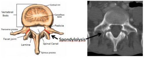
Another term you may see with regard to the pars is spondylolysis. This is defined as a defect in the pars interarticularis, typically from a fracture. So if you see either of these terms in a radiology report, recognize that they are basically one and the same.

Here is a nice image showing the location of the pars, and the axial CT appearance of “bilateral pars defects.”

Mystery solved! Amaze your friends!

Print Friendly, PDF & Email

The Cost Of Duplicate Radiographic Studies

Speaking of radiation, here’s another tidbit. Duplicate radiographic studies are a continuing issue for trauma professionals, particularly after transfer from a smaller hospital to a trauma center. The incidence has been estimated anywhere from 25% to 60% of patients. A lot has been written about the radiation dangers, but what about cost?

A Level II trauma center reviewed their experience with duplicate studies in orthopedic transfer patients retrospectively over a one year period. They looked at the usual demographics, but also included payor, cost information, and reason for repeat imaging. Radiation dose information was also collected.

Here are the factoids:

  • 513 patients were accepted from 36 referring hospitals
  • 48% had at least one study repeated, 256 CT scans and 161 conventional imaging studies
  • Older patients and patients with low GCS were much more likely to receive repeat studies
  • There were no association with the size of the referring hospital or the ability of the patient to pay
  • Most transfers had commercial insurance; only 11% had Medicaid and 17% were uninsured
  • Additional radiation from repeat scans was 8 mSv. The average radiation dose from both hospitals was 38 mSv. This is 13 years of background radiation exposure!
  • The cost of all the repeat studies was over $96,000

Bottom line: This is an eye-opening study, particularly regarding how often repeat imaging is needed, how much additional radiation is delivered, and now, the cost. And remember that these are orthopedic patients, many of whom had isolated bony injuries. I would expect that patients with multiple and multi-system injuries would require more repeat imaging and waste even more money. It is imperative that all centers that receive transfers look at adopting some kind of electronic data transfer for imaging, be it a VPN or some cloud-based service. With the implementation of the Orange Book by the American College of Surgeons, Level I and II centers will receive a deficiency if they do not have some reliable mechanism for this.

“Level I and II facilities must have a mechanism in place to view radiographic imaging from referring hospitals within their catchment area (CD 11–42).”

Reference: Clinical and Economic Impact of Duplicated Radiographic Studies in Trauma Patients Transferred to a Regional Trauma Center. J Ortho Trauma 29(7):e214-e218, 2015.

Print Friendly, PDF & Email

How Much Radiation Exposure In Imaging Studies?

Everyone knows that CT scans deliver more radiation than conventional x-ray. But how much does each test really deliver? And how significant is that?

Let me try to put it all into perspective. First, how much radiation are we exposed to just living outside the hospital? Background radiation is everywhere. It consists of radioactive gases (argon) in the air we breathe, radiation from the rocks and other things around us, and cosmic rays blasting through us from space.

In the United States, the average background radiation each of us is exposed to is about 3.1 milliSieverts (mSv). I’ve compiled a table to show the approximate dose delivered by some of the common radiographic studies ordered by trauma professionals. And to keep it real, I’ve calculated how much extra background radiation we would have to absorb, in units of time, to have an equivalent exposure.

Read and enjoy! Remember, doses may vary by scanner, settings, and dose reduction measures used.

Test Dose (mSv) Equivalent background
radiation
Chest x-ray 0.1 10 days
Pelvis x-ray 0.1 10 days
CT head 2 8 months
CT cervical spine 3 1 year
Plain c-spine 0.2 3 weeks
CT chest 7 2 years
CT abdomen/pelvis 10 3 years
CT T&L spine 7 2 years
Plain T&L spine 3 1 year
Millimeter wave
scanner (that hands
in the air TSA thing at
the airport)
0.0001 15 minutes
Scatter from a chest
x-ray in trauma bay
when standing one 
meter from the
patient
0.0002 45 minutes
Scatter from a chest
x-ray in trauma bay
when standing three 
meters from the
patient
0.000022 6 minutes
Print Friendly, PDF & Email

How Much Radiation is the Trauma Team Really Exposed To?

Previously, I posted about “other people” wearing perfectly good lead aprons lifting them up to their chin during portable xrays in the trauma bay. Is that really necessary, or is it just an urban legend?

Lead apron fly

After hitting the medical radiation physics books (really light reading, I must say), I’ve finally got an answer. Let’s say that the xray is taken in the “usual fashion”:

  • Portable technique in your trauma bay
  • Tube is approximately 5 feet above the xray plate
  • Typical chest settings of 85kVp, 2mAs, 3mm Al filtration
  • Xray plate is 35x43cm

The calculated exposure to the patient is 52 microGrays. Most of the radiation goes through the patient onto the plate. A very small amount reflects off their bones and the table itself. This is the scatter we worry about.

So let’s assume that the closest person to the patient is 3 feet away (1 meter). Remember that radiation intensity diminishes as the square of the distance. So if the distance doubles, the intensity decreases to one fourth. By calculating the intensity of the small amount of scatter at 3 feet from the patient, we come up with a whopping 0.2 microGrays. Since most people are even further away, the dose is much, much less for them.

Let’s put it perspective now. The background radiation we are exposed to every day (from cosmic rays, brick buildings, etc) amounts to about 2400 microGrays per year. So 0.2 microGrays from chest xray scatter is less than the radiation we are exposed to naturally in about 44 minutes!

The bottom line: unless you need to work out you shoulders and pecs, don’t bother to lift your lead apron every time the portable xray unit beeps. It’s a waste of time and effort, unless you are dealing with xray imaging on a very regular basis! And that 52 microGrays the patient absorbed? That’s 8 days worth of background radiation.

Print Friendly, PDF & Email

Wear The Damn Lead Gown!

Pet peeve time. All trauma team members must wear personal protective equipment (PPE) when they attend a trauma activation. It’s for their own protection as well as our patients’. When I am the primary faculty at any trauma activation, I quickly scan all the other team members to ensure they are wearing it. If not, I give them a “gentle reminder” that they need to go get dressed properly.

This is all well and good. But recently I’ve noticed a trend when it comes time to shoot the basic x-rays needed for assessment (chest and/or pelvic images). When the radiology tech calls out to clear the torso and make sure someone else’s head or hands are not over the patient, half the team goes running out of the room. They are missing one key component of their gear:

Yes, their lead gown! Now granted, the amount of radiation exposure is not huge as I’ve documented in previous posts. But it is cumulative and for safety reasons, x-ray exposure must be limited.

But is running out of room the best way to decrease exposure? I think not! This is very disruptive to the way the team should function and interrupts patient care. Ideally, everyone in the room within 2-3 meters of the x-ray tube should be shielded in some way. And the most effective way to do this is to wear the damn lead gown!

Bottom line: I’ve adjusted my scan when the trauma team assembles. I now look for a lead gown underneath the usual PPEs. And if I don’t see it, I remind the offenders that, if they leave the room when the x-rays are taken, I’m not letting them back in. It’s been very effective at reversing this troubling trend.

In my next two posts, I’ll detail how much radiation the team is exposed to, and how much our patients receive from the studies we order.

Print Friendly, PDF & Email