Category Archives: Imaging

Duplex Ultrasound For DVT: How Does It Work?

Admit it. You’re curious. You order this test for your trauma patients all the time but you’ve never seen it done. It’s simple and noninvasive, but it does require access to all areas to be evaluated. This means that extremities that are casted or splinted, or that have extensive dressings in place may be incompletely evaluated.

The study is called “duplex” because it makes use of two modalities: traditional ultrasound and Doppler ultrasound. Traditional ultrasound is used to view the compressibility of the veins of interest at a number of locations. Doppler measures the speed of blood flow under the probe, and can show areas of sluggish flow.

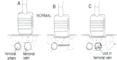
The following diagram shows the traditional ultrasound technique being used to compress the vein of interest (femoral, popliteal, etc.). Part A shows the probe gently resting over the vessels. Part B shows a fully compressible vein (normal), and Part C shoes partial compression due to the presence of thrombus.

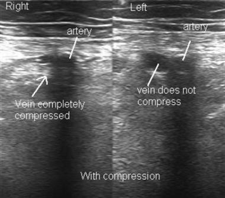
The following diagram shows what the actual ultrasound study looks like. The right side is normal, but the left side shows a venous thrombosis.

EAST 2017 #6: FAST Exam After Rolling to the Right

The FAST exam is an integral part of trauma evaluation. Even after experience and credentialing of providers, there tends to be some variability in performance. This is especially true when the abnormal findings (or amount of fluid present) is relatively small.

Can we improve this by doing something as simple as using gravity to help? When the patient is supine, fluid tends to pool in the pelvis, where interpretation is a little more complicated.  The surgery program at Guthrie/Packer Hospital created a small pilot study to see if they might improve the sensitivity of FAST by rolling patients to their right briefly, before returning to the supine position and performing the exam.

They enrolled seven participants who were already undergoing peritoneal dialysis (PD), so there was easy access to the peritoneal cavity for administration of known amounts of free fluid. First, each patient was drained of any residual dialysate via their PD catheter. They then underwent a baseline FAST exam. Next, they were placed in the right lateral decubitus position for 30 seconds, then placed supine again and the FAST was repeated. Each patient then had 50cc of dialysate infused, and the process was repeated until a positive FAST was obtained.

Here are the factoids:

  • Of the seven patients recruited, one was excluded because the initial FAST was equivocal due to body habitus and polycystic kidney disease
  • A maximum of 3 aliquots were given (150cc max)
  • Two patients became positive after right side down before any additional fluid was infused
  • None of the four remaining patients had a positive FAST after infusion of any aliquot in the supine position
  • All four became positive after the right side down maneuver,  two after 50cc, one after 100cc, and one after 150cc

Bottom line: The authors conclude that this may be a valuable technique to help detect smaller quantities of fluid than we normally do. I’m not so sure. First, it’s a tiny study in a patient group that is very different from trauma. And it’s impossible to quantify how much dialysate was left after initial drainage of the PD catheter. Finally, we know that FAST can’t “see” small quantities of fluid, but we have constructed our management algorithms around this fact. So we have a good idea of when we should do further imaging or run off to the operating room. Making this test more sensitive may skew these practice guidelines toward doing more (and potentially unneeded) imaging and surgery.

Questions and comments for the authors/presenters:

  • Did you record the volumes and administration times of dialysate given prior to the study? This may correlate with the initial positives and volumes needed to give a positive result.
  • Similarly, did you look at BMI and body habitus to see if there might be a correlation?
  • Are you planning any type of followup study, as you suggested in the abstract?

Click here to go the the EAST 2017 page to see comments on other abstracts.

Related posts:

Reference: Can we be faster? FAST examination after rolling to the right dramatically increases sensitivity. Quick Shot #7, EAST 2017.

EAST 2017 #5: Subarachnoid Hemorrhage, Neurosurgical Consults, and Repeat Head CT

Neurosurgical involvement in the management of simple traumatic brain injury (TBI) has been slowly dwindling over the past several years. This is the result of the general consensus that very few of these patients progress to need neurosurgical procedures.

A group at Wright State University in Dayton sought to define the progression of one specific finding in TBI, the subarachnoid hemorrhage (SAH). Secondarily, the wanted to determine if a neurosurgery consultation was warranted in these patients.

They performed a five year retrospective review of their registry data, identifying patients with both mild TBI (GCS 13-15) and SAH. They excluded patients with any other brain lesion on CT.

Here are the factoids:

  • 301 patients were enrolled during the 5 year period
  • All had a neurosurgical consultation
  • Time between the initial CT and a followup scan was about 11 hours
  • 91% showed stable or resolving SAH on the followup scan
  • 9% showed a worsening SAH or additional lesions on the repeat scan

Bottom line: The authors conclude that initial neurosurgical consultation is not needed, since only 9% of patients have worrisome findings on repeat CT. They do, however, recommend that the practice of repeat scanning be continued because of this same number.

Our trauma service looked at this issue a year ago, and determined that most of these lesions either do not progress, or never require any intervention if they do, with a few notable exceptions. For that reason, we abandoned both neurosurgical consultation and repeat CT scans for patients with non-aneurysmal SAH, a single parenchymal hemorrhage, or linear skull fractures. We continue to do both for patients with epidural and/or subdural hemorrhage. You can download a copy of this protocol here.

Questions and comments for the authors/presenters:

  • Did you look at platelet count or INR in the study. Were patients excluded based on abnormal values?
  • Did every patient get a repeat scan?
  • Break down the lesions in the 9% of patients who had some sort of progression or new finding. Did you see any common themes (age, chronic alcohol use, etc.)?
  • Did you encounter any patients with “non-central SAH”, that might indicate an aneurysm? How were they dealt with?
  • How has or will your trauma service change its practice based on your findings.

Click here to go the the EAST 2017 page to see comments on other abstracts.

Related posts:

Reference:   Management of subarachnoid hemorrhage (SAH) by the trauma service: are repeat CT scanning & routine neurosurgical consultation necessary? Poster #16, EAST 2017.
EAST 2017 #2: CT Scan After Recent Operative Exploration for Penetrating Trauma

The general rule for penetrating trauma, especially gunshots to the abdomen, is that you don’t need to obtain a CT scan to help you decide to go to the OR. (Of course, there are a few exceptions.) And the corollary has always been that you don’t need to get a CT scan after you operate for penetrating trauma.

But the group at UCSF is questioning this. They retrospectively looked at 5 years of data on patients who underwent trauma laparotomy without preoperative imaging. They focused on new findings on CT that were not reported during the initial operation.

Here are the factoids:

  • 230 of 328 patients undergoing a trauma lap did not have preop imaging
  • 85 of the 230 patients (37%) underwent immediate postop CT scan. These patients tended to have a gunshot mechanism and higher injury severity score.
  • Unreported injuries were found in 45% (!) and tended to be GU and orthopedic in nature
  • 47% of those with unreported injuries found required some sort of intervention

Bottom line: This is a very interesting and potentially practice changing study. However, there is some opportunity for bias since only select patients underwent postop scanning. Nevertheless, one in five patients who did get a postop scan had an injury that required some sort of intervention. This study begs to be reworked to further support it, and to develop specific criteria for postop scanning.

Questions/comments for the authors/presenters:

  • Be sure to break down your results by gunshot vs stab. This will help formulate those criteria I mentioned above.
  • Specifically list the occult injuries and interventions required. In some studies, those “required interventions” are pretty weak (urology consult vs an actual procedure).
  • How exactly did the operating surgeons determine who to send to CT? Was it surgeon-specific (i.e. one surgeon always did, another never did)? Was it due to operative findings (hole near the kidney)? This is also needed when developing specific criteria for postop imaging.
  • Nice poster!

Click here to go the the EAST 2017 page to see comments on other abstracts.

Related posts:

Reference: Routine tomography after recent operative exploration for penetrating trauma: what injuries do we miss?  Poster #14, EAST 2017.

EAST 2017 #1: Accuracy of CT Scans Done Outside The Trauma Center

Imaging prior to transfer to a trauma center has been the subject of debate for years. The focus has primarily been on the necessity of these scans, and the sheer numbers that are done. For the most part, the discussion has been driven by the potential for radiation exposure.

This paper, from the University of Oklahoma, takes a different approach. The authors looked at the accuracy and adequacy of imaging performed prior to transfer to their Level I trauma center.

Patients were enrolled prospectively over an 8 month period in 2012. Outside images were interpreted by a single radiologist who was blinded to the outside interpretation.  If images were repeated, they were compared to the first scan, and the reason for the redo was noted.

Here are the factoids:

  • 235 consecutive transfer patients were enrolled, and 203 who had at least one CT scan were included in the final dataset
  • 76% of these patients had additional imaging performed once they arrived at the trauma center
  • Reasons for additional images were insufficient workup (76%) and technical inadequacy (31%)
  • Missed injuries were detected on outside CT scans 49% of the time, and the majority of them (90%) were deemed clinically significant
  • Missed injuries on a repeated scan were present in 54% of patients, and 76% were clinically significant
  • Overall, 73% of images (either outside or repeat) contained additional injuries

Bottom line: This is a new approach to assessing the value of outside imaging prior to transfer to a trauma center. I have always recommended that trauma centers work with their referral partners to assure them we don’t need them to do the workup for us. I encourage them to obtain only what they need to decide if they can keep the patient. But once they find anything that they cannot treat, stop all workup and prepare to transfer.

Questions/comments for the authors/presenters:

  • Why did you use such an old dataset?
  • Is this a prospective enrollment/retrospective analysis study designed to use an existing, old dataset?
  • How did you decide that outside imaging represented an inadequate workup? Do you have a diagnostic imaging guideline that you follow?
  • What are the credentials for your trauma radiologist?
  • How did you determine that a missed injury was clinically significant? Be sure to provide a list and explanation during your presentation.
  • Be sure to separate out missed injuries seen on the original CT from new missed injuries seen on the repeat scan.
  • Congratulations on looking at an old problem in a new way!

Click here to go the the EAST 2017 page to see comments on other abstracts.

Related posts:

Reference: Adequacy and accuracy of non-tertiary trauma center computed tomography: what are we missing? Paper #7, EAST 2017.