Category Archives: Imaging

Metal Splints – Can You X-ray Through Them?

Splinting is an important part of the trauma resuscitation process. No patient should leave your trauma resuscitation room without splinting of all major fractures. It reduces pain, bleeding, and soft tissue injury, and can keep a closed fracture from becoming an open one.

But what about imaging? Can’t the splint degrade x-rays and hamper interpretation of the fracture images? Especially those pre-formed aluminum ones with the holes in them? It’s metal, after all.

Some of my orthopedic colleagues insist that the splint be removed in the x-ray department before obtaining images. And who ends up doing it? The poor radiographic tech, who has no training in fracture immobilization and can’t provide additional pain control on their own.

But does it really make a difference? Judge for yourself. Here are some knee images with one of these splints on:
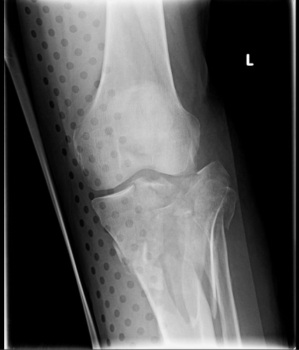
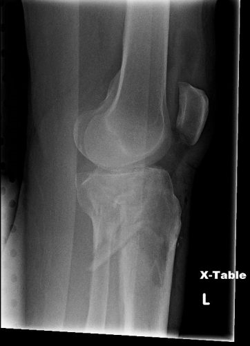
Amazingly, this thin aluminum shows up only faintly. There is minimal impact on interpretation of the tibial plateau. And on the lateral view, the splint is well posterior to bones.
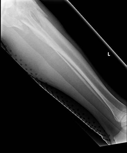
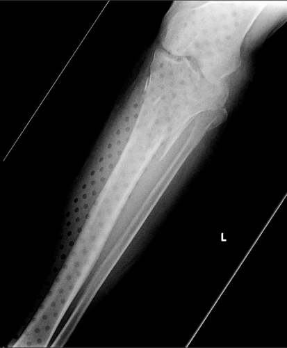
On the tib-fib above, the holes are a little distracting on the AP view, but still allow for good images to be obtained.

Bottom line: In general, splints should not be removed during the imaging process for acute trauma. For most fractures, the images obtained are more than adequate to define the injury and formulate a treatment plan. If the fracture pattern is complex, it may be helpful to temporarily remove it, but this should only be done by a physician who can ensure the fracture site is handled properly. In some cases, CT scan may be more helpful and does not require splint removal. And in all cases, the splint should also be replaced immediately at the end of the study.


Print Friendly, PDF & Email

Don’t Repeat Trauma Activation X-rays!!

You are in the middle of a fast-paced trauma activation. The patient is awake, and mostly cooperative. The x-ray plate is under the patient and everyone stands back as the tech gets ready to fire the x-ray machine. At that very moment, your patient reaches up and places his hand on his chest. Or one of the nurses reaches over to check an IV site.

The x-ray tech swears, and offers to re-shoot the image. What do you do? Is it really ruined? They have an extra plate in hand and are ready to slide it under the patient bed.

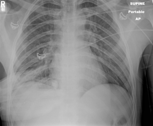
The decision tree on this one is very simple. There are two factors in play: what do you need to see, and how hard is it to see? The natural reaction is to discard the original image and immediately get a new one. It’s so easy! Plus, the techs will take heat from the radiologist because of the suboptimal image. But take a look at this example of a “ruined” chest xray.

It’s just the patient’s hand! You can still see everything that you really need to.

Bottom line: You are looking for 2 main things on the chest x-ray: big air and big blood. Only those will change your management in the trauma bay. And they are very easy to see. Couple that with the fact that an arm overlying the image does not add a lot of “noise” to the image. So look at the processed image first. 99% of the time, you can see what you need, and will almost never have to repeat. [Hint: the same holds true for the pelvic x-ray, too. You are mainly looking for significant bony displacements, which are also easy to see.]

Related posts:

Print Friendly, PDF & Email

Do We Need Cervical MRI Scans If The CT Is Negative?

The debate on how to clear the cervical spine just never ends. We have finally come to some degree of agreement that certain patients (awake, alert, not impaired or head injured, without distracting injury) can undergo clinical clearance alone.

But if those criteria are not met, what next? Universally, adults receive a CT scan of the cervical spine. In the majority of centers, this is coupled with a good clinical examination. And if both are negative, the collar can be removed.

But recent literature suggest that a good, high-quality cervical CT read by a skilled neuroradiologist may be good enough. This has been demonstrated in several papers involving patients who are comatose or other-wise unable to participate with a clinical exam.

Many centers and trauma professionals are still reluctant to remove the cervical collar without that clinical examination. A new study asked the question: would an MRI provide additional, significant information over and above the CT scan in those patients who could not be examined or had persistent neck pain?

A consortium of 8 Level I and II trauma centers in New England participated in this study coordinated by Yale. Blunt trauma patients who underwent MRI after negative cervical CT were considered for the study. On further review, if they received the scan because they could not be clinically evaluated, or if they had complaints of persistent neck pain, they were enrolled. CT scanners with at least 64-slice capabilities were required. There was no mention of the qualifications or special experience of the radiologists reading the images at each center.

Here are the factoids:

  • 767 patients were enrolled in this 30-month study. A total of 43% were for persistent neck pain, 44% for inability to examine, and 9% for both.
  • Nearly a quarter had an abnormal MRI scan:
    • 17% ligamentous injury
    • 4% soft tissue swelling
    • 1% disk injury
    • 1% dural hematoma
  • The collar was removed in most (88%) patients with a normal MRI, but in only 13% with ab-normal MRI
  • 11 patients underwent a surgical procedure and half had neurologic signs or symptoms. 10 of them had ligamentous injury, 1 had dural hematoma, and 1 had both

Bottom line: Looks almost compelling, right? One would think that we had better get an MRI on all of these patients! But read more closely, please. Yes, injuries were found. But did they really “require” an intervention? For some injuries, it’s a chip shot. A three column ligamentous injury equals stabilization in any textbook. But management of lesser injuries is less clear. And could some of these injuries have been recognized by a skilled neuroradiologist reading the CT image?

So what to do? There is not enough data for a universal protocol yet. Unfortunately, you will need to develop your own institutional policy based on the experience and opinions of your spine and neurosurgeons. They are the ones who will have to deal with the decision making during and after these studies. Until the definitive study comes along.

Reference: Cervical spine MRI in patients with negative CT: A prospective, multicenter study of the Research Consortium of New England Centers for Trauma (ReCONECT). J Trauma 82(2):263-269, 2017.

Print Friendly, PDF & Email

Duplex Ultrasound For DVT: How Does It Work?

Admit it. You’re curious. You order this test for your trauma patients all the time but you’ve never seen it done. It’s simple and noninvasive, but it does require access to all areas to be evaluated. This means that extremities that are casted or splinted, or that have extensive dressings in place may be incompletely evaluated.

The study is called “duplex” because it makes use of two modalities: traditional ultrasound and Doppler ultrasound. Traditional ultrasound is used to view the compressibility of the veins of interest at a number of locations. Doppler measures the speed of blood flow under the probe, and can show areas of sluggish flow.

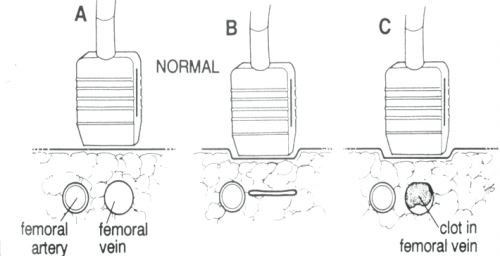
The following diagram shows the traditional ultrasound technique being used to compress the vein of interest (femoral, popliteal, etc.). Part A shows the probe gently resting over the vessels. Part B shows a fully compressible vein (normal), and Part C shoes partial compression due to the presence of thrombus.

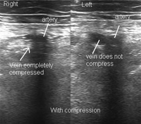
The following diagram shows what the actual ultrasound study looks like. The right side is normal, but the left side shows a venous thrombosis.

Print Friendly, PDF & Email