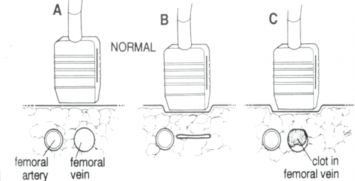
Tag Archives: ultrasound

Best Of EAST #5: Ultrasound vs Chest X-Ray After Chest Tube Removal

The chest is one of the most commonly injured body regions. Patients are frequently found to have either air or blood in the chest, and many require a chest tube (tube thoracostomy) for these conditions. There is an art to chest tube removal, and even in 2021, the best practice has not been fully worked out.

Some believe that pulling the tube during a breath hold is best. Others do this during full expiration. Most centers confirm an uneventful tube removal with a plain chest x-ray. But the time interval after removal varies considerably.

The trauma group at the University of Tennessee – Chattanooga examined the use of chest ultrasound as the confirmatory test for residual pneumothorax after removing a chest tube. They developed an institutional practice guideline requiring a trans-thoracic ultrasound performed by a first-year resident two hours after tube removal. The interns all completed a 30-minute standard ultrasound course for training prior to beginning the study.

Two hours after tube removal, an intern performed the ultrasound (US) and interpreted it. A chest x-ray (CXR) was then ordered and the results compared.

Here are the factoids:

  • A retrospective review of 46 patients was performed, but the inclusion criteria were not listed in the abstract
  • Eleven of the 46 (24%) had a residual pneumothorax on CXR, and the US detected it in 12 (26%)
  • Three patients had PTX on CXR, but not US
  • Four patients had PTX on US, but not CXR
  • None of the PTX were clinically significant, and none required tube reinsertion
  • Cost of care savings was projected to be $4,000 if chest x-ray was not needed

The authors concluded that bedside ultrasound was an acceptable alternative to chest x-ray, with decreased radiation exposure and cost.

Bottom line: This is an intriguing abstract. It shows us that there might be an alternative to the standard chest x-ray confirmation after chest tube removal. It’s a very small study, so non-inferiority can’t truly be established yet. The studies are complementary since each study misses a few pneumothoraces that the other picks up.

At this point, I wouldn’t recommend switching entirely to ultrasound until we have a larger series. But I bet we will be able to in the future. Ultimately, this could reduce radiation exposure (tiny anyway for a chest x-ray) and save a small amount of money. But it will reduce x-ray department resource usage, which may be very helpful for the hospital.

Here are my questions for the authors and presenter:

  • How did you select your patients? What were the selection criteria? How long did it take to accrue 46 patients? It’s important that all patients with a chest tube had the criteria applied, otherwise there is an opportunity for bias. We want to make sure that you didn’t inadvertently enroll only the patients for whom ultrasound works well.
  • How much of a burden was placed on the interns who did the exam? Was the ultrasound unit nearby? Or did they have to spend 30 valuable minutes rolling it to the floor and doing the study? Radiology department resource use needs to be balanced with intern resource utilization.
  • Why did you have such a high rate of residual pneumothorax after the tubes were pulled (about 25%). This seems a bit higher than what the literature reports.
  • What does your protocol require when a residual pneumothorax is found? Do you have to perform another study after an additional time interval to prove that it is not getting larger? Serial ultrasound exams? Another chest x-ray? Please show us your entire guideline.

I really enjoyed this paper. I’m looking forward to hearing the nitty gritty details during the presentation.

Reference: ULTRASOUND SAFELY REPLACES CHEST RADIOGRAPH AFTER TUBE THORACOSTOMY REMOVAL IN TRAUMA PATIENTS. EAST 25th ASA, oral abstract #9.

Duplex Ultrasound For DVT: How Does It Work?

Admit it. You’re curious. You order this test for your trauma patients all the time but you’ve never seen it done. It’s simple and noninvasive, but it does require access to all areas to be evaluated. This means that extremities that are casted or splinted, or that have extensive dressings in place may be incompletely evaluated.

The study is called “duplex” because it makes use of two modalities: traditional ultrasound and Doppler ultrasound. Traditional ultrasound is used to view the compressibility of the veins of interest at a number of locations. Doppler measures the speed of blood flow under the probe, and can show areas of sluggish flow.

The following diagram shows the traditional ultrasound technique being used to compress the vein of interest (femoral, popliteal, etc.). Part A shows the probe gently resting over the vessels. Part B shows a fully compressible vein (normal), and Part C shoes partial compression due to the presence of thrombus.

The following diagram shows what the actual ultrasound study looks like. The right side is normal, but the left side shows a venous thrombosis.

EAST 2016: Measuring Volume Status Using Jugular Ultrasound

We’re getting pretty handy using ultrasound at
the bedside to tell us some interesting things. It started with FAST exams in
the ED. Then we added a few views and came up with the Extended FAST, which was
helpful in showing potential chest pathology.

Ultrasound made its way into other areas of the
hospital, and is now used routinely to place IV lines, arterial lines, and
central venous catheters. I’ve previously written about using ultrasound to
evaluate volume status by imaging the IVC in the abdomen. And now, the group at
Shock Trauma in Baltimore is trying to reach even further.

They are now using IVC variations and cardiac
stroke volume variations to assist in volume assessment in critically ill
patients. These studies have a learning curve, especially the stroke volume
calculations. They performed a study that evaluated another possible window
into the patient’s volume status, the positional internal jugular change.

The diameter of the IJ was evaluated while the patient
was flat, and again when the head was elevated to 90 degrees. A fluid bolus was
given, and the positional change in diameter was measured again. The results
were then correlated with changes in measured stroke volume of at least 10%.

Here are the factoids:

  • This prospective, observational study involved 159 patients over 1.5
    years
  • Positional IJ diameter change was much better than IVC diameter changes
    (receiver operating characteristic areas of 0.93 vs 0.67)
  • The authors tried to use the stroke volume variation during passive leg
    raise (odd, but doesn’t involve sitting the patient up), and concluded they
    could not accurately assess it. This arm of the study was abandoned.

Bottom
line: Leave it to the folks at Shock Trauma to come up with more weird yet interesting
stuff. This is very preliminary data, and their analysis is ongoing. Any
application of this study will be somewhat limited, since many patients are not
allowed to sit up due to their injuries or baseline hemodynamic status. We will
see where this technique
ends up: in our armamentarium, or in the trash heap.

Go for the
jugular: assessing volume responsiveness in critically ill surgical patients.
EAST 2016 Oral abstract #32.

FAST Cardiac Ultrasound And Traumatic Arrest

Cardiac arrest in trauma patients is bad. Really bad. There are few survivors, mainly those who have some signs of life when they roll into the resuscitation room. One of the signs we look for is cardiac electrical activity, especially a narrow complex rhythm. But most of the time these patients don’t survive either. Could there be a way to fine tune the use of pulseless electrical activity (PEA) to better determine when further care is futile?

The trauma group at UCSF-East Bay did a nice, retrospective review on the use of the cardiac portion of the FAST exam to assess patients arriving in PEA arrest after either blunt or penetrating trauma. The numbers were a bit thin, but they were able to study 162 patients who had both FAST and EKG upon arrival. Of those patients, 71 had electrical activity, but only 17 had cardiac motion. However, 4 of these 17 survived (24%) vs only 1 of the 54 who did not have cardiac motion.

About a third of these 71 patients suffered blunt trauma, the remainder had penetrating injury. Of the 17 with cardiac activity, 14 were penetrating and 3 were blunt. And of the 4 survivors mentioned above, 3 were penetrating.

Only 1 of the 71 patients with PEA and no cardiac activity survived, and this was a blunt arrest(!).

Bottom line: Traumatic arrest is a generally fatal problem. However, it appears that use of the cardiac portion of the FAST exam in penetrating or blunt trauma can help fine tune the aggressiveness of resuscitation. PEA without cardiac activity is uniformly fatal (although there was one blunt survivor, the authors did specify the quality of this survival). It may be wise to forego further resuscitative efforts in PEA patients without cardiac activity because they will not survive, even as an organ donor.

Reference: The heart of the matter: Utility of ultrasound of cardiac activity during traumatic arrest. J Trauma 73(1):103-110, 2012.