Tag Archives: Penetrating trauma

The Role Of Postop CT Scan In Penetrating Trauma

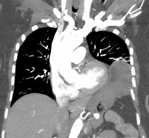
CT scans are commonly used to aid the workup of patients with blunt trauma. They are occasionally useful in penetrating trauma, specifically when penetration into a body cavity is uncertain and the patient has no hard signs that would send him or her immediately to the operating room.

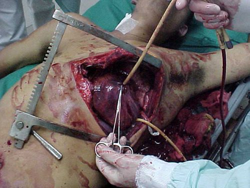
Is there any role in operative penetrating trauma, after the patient has already been to the OR? The dogma has always been that the eyeballs of the surgeon in the OR are better than any other imaging modality. Really? The surgical group at San Francisco General addressed this question by retrospectively reviewing 6 years of their operative penetrating injury registry data. They were interested in finding how many occult injuries (seen with CT but not by the surgeon) were found on a postop CT. A total of 225 patients who underwent operative management of penetrating abdomen or chest injury were included. Here are the factoids:

  • Only 110 patients had a postop CT scan; 73 had scans within the first 24 hours, the other 37 were scanned later
  • Rationale for early scan was to investigate retroperitoneal injury in half of patients, but frequently no indication was given (41%)
  • Rationale for late scan was for workup of ileus in one third, or for evaluation of new or unexpected clinical problems
  • Occult injuries were found in about half of early CT patients (52%), and 22% of late CT patients
  • The most common occult injuries were fractures, GU issues, regraded solid organ injury, and unrecognized vascular injuries
  • Ten patients had management changes, including:
    • Interventional radiology for four injuries with extravasation
    • Operation for orthopedic or GU injury in seven patients
    • One patient underwent surgery for an unstable spine fracture

Bottom line: There appears to be a significant benefit to sending some penetrating injury patients to CT in the early postop period. Specifically, those with injury to the retroperitoneum, deep into the liver, near the spine, or with multiple and complicated injuries would benefit. Simple stabs and gunshots that stay away from these areas/structures probably do not need followup imaging. 

Reference: Routine computed tomography after recent operative exploration for penetrating trauma: What injuries do we miss? J Trauma 83(4):575-578, 2017.

What Is: A Rubber Bullet?

The protests in cities across the country continue. Many are peaceful, but not all. In some cases, police have resorted to “non-lethal” weapons to control and disperse crowds.

Although these weapons are called non-lethal, that’s not entirely true. The projectiles, gases, and powders that are being used all have some degree of morbidity and mortality. They are certainly less so than traditional projectiles (bullets), but serious and fatal injures can and do occur.

One item that is talked about in the news is the rubber bullet. What are these, exactly? The generic term is a “kinetic impact projectile” (KIP). It encompasses a variety of objects that are not designed to penetrate flesh like a regular bullet. They can be bullets, beanbags, sponges, pellets, and other odds and ends.

And the so-called “rubber bullet” isn’t even necessarily made of rubber. It can be plastic, metal, rubber, or other substances.

There is very little published data on injuries caused by KIPs. Because of their odd shapes, they tend to tumble when they are fired. This decreases aiming accuracy substantially when the target is distant. They are designed to be aimed at the lower extremities. However, if the aim is too high or the round is fired at close range, it can be lethal.

Here are some typical injuries that have been descirbed:

  • Subdural and intraparenchymal hematomas
  • Skull and facial fractures
  • Eye injuries leading to blindness (this happened to a photographer in Minneapolis last week)
  • Rib fractures and pulmonary contusions
  • Spleen laceration
  • Blunt intestinal injury

Here’s a video from one of the manufacturers that shows the amount of target deformation caused by a sponge tipped bullet. Very impressive.

YouTube player

(Tumblr viewers please click here to view video)

Bottom line: Although they sound relatively innocuous, kinetic impact projectiles of any kind are far from it. If you are called to treat a patient who has been shot with one, be sure to do a very thorough evaluation. Most head, neck, and torso injuries should undergo CT scanning to delineate deep or occult injuries in detail. In-hospital observation of torso injuries is warranted, as well as a good tertiary exam.

Off-Label Use of the Foley (Urinary) Catheter

Foley catheters are a mainstay of medical care in patients who need control or measurement of urine output. Leave it to trauma surgeons to find warped, new ways to use them!

Use of these catheters to tamponade penetrating cardiac injuries has been recognized for decades (see picture, 2 holes = 2 catheters!). Less well appreciated is their use to stop bleeding from other penetrating wounds.

Foley catheters can be inserted into just about any small penetrating wound with bleeding that does not respond to direct pressure. (Remember, direct pressure is applied by one or two fingers only, with no flat dressings underneath to diffuse the pressure). Arterial bleeding, venous bleeding or both can be controlled with this technique.

In general, the largest catheter with the largest possible balloon should be selected. It is then inserted directly into the wound until the entire balloon is inside the body. Inflate the balloon using saline until firm resistance is encounted, and the bleeding hopefully stops. Important: be sure to clamp the end of the catheter so the bleeding doesn’t find the easy way out!

Use of catheter tamponade buys some time, but these patients need to be in the OR. In general, once other life threatening issues are dealt with in the resuscitation room, the patient should be moved directly to the operating room. In rare cases, an angiogram may be needed to help determine the type of repair. However, in the vast majority of cases, the surgeon will know exactly where the injury is and further study is not needed. The catheter is then prepped along with most of the patient so that the operative repair can be completed.

Tomorrow: an off-label use for this catheter in abdominal trauma!

How To Evaluate A Stab To The Diaphragm – Part 1

Penetrating injury to the diaphragm, and specifically stab wounds, have been notoriously hard to diagnose since just about forever. Way back in the day (before CT), we tried all kinds of interesting things to help figure out if the patient had a real injury. Of course, we could just go to the OR and lap the patient (laparoscopy did not exist then). But the negative lap rate was significant, so we tried a host of less invasive techniques.

Remember diagnostic peritoneal lavage? Yeah, we tried that. The problem was that the threshold for red cells per cubic mm was not well defined. Some would supplement this technique with a chest tube to see if lavage fluid would drain out. And one paper described instilling nuclear medicine tracer into the abdomen and sitting the patient under a gamma camera for a few hours to see if any ended up in the chest. Groan!

We thought that CT would save us. Unfortunately, resolution was terrible in the early years. If you could actually see the injury on CT, it was probably because a large piece of stomach or colon had already fallen through it. But as detectors multiplied and resolution improved, we could begin to see some smaller defects. But we still missed a few. And the problem is that left-sided diaphragmatic holes slowly enlarge over time (years), until the stomach or colon falls through it. (See below)

A group of radiologists and surgeons in a Turkish trauma hospital recently published a modest series of patients with left-sided diaphragm injuries evaluated by CT. They looked at about 5 years of their experience in a group of patient who were at risk for the injury due to a thoraco-abdominal stab wound. Unstable patients were immediately taken to OR. All of the remaining patients underwent an initial CT scan, followed by diagnostic laparoscopy after 48 hours if they remained symptom free.

Here are the factoids:

  • A total of 43 stable patients with a left thoraco-abdominal stab were evaluated
  • 30 patients had a normal CT, and 13 had the appearance of an injury
  • Of those who were CT positive, only 9 of 13 (69%) actually had the injury at operation
  • Two of the 30 (7%) who were CT negative were found to have a diaphragm injury during followup laparoscopy
  • So in the author’s hands, there was 82% sensitivity, 88% specificity, a positive predictive value of only  69%, and a negative predictive value of 93%

Bottom line: The authors somehow looked at the numbers and concluded that CT is valuable for detecting left diaphragm injury. Huh? They missed 7% of injuries, only finding them later at laparoscopy. And they had a 31% negative laparotomy rate. 

Now, it could be that the authors were using crappy equipment. Nowhere in their paper do they state how many detectors, or what technique was used. Since it took place over a 5 year period, it is quite possible that the earlier years of the study used equipment now considered to be out of date, or that there was no standardized technique.

CT may not yet be ready for prime time. But it can be a valuable tool. Tune in tomorrow for some tips on how and when to look for this insidious injury.

Reference: Evaluation of diaphragm in penetrating left thoracoabdominal
stab injuries: The role of multislice computed tomography. Injury 46:1734-1737, 2015.

The “Egg Timer” Injury

Most patients with major traumatic injuries are handled in a very systematic way by both EMS and trauma centers. We have routines and protocols designed to provide rapid, quality care to these individuals. But over the years, I’ve begun to appreciate the fact that there is a very small subset of these patients who are different.

I term these patients as having an “egg timer injury”. These are patients who have only a certain number of minutes to live. This fact requires us to change the usual way we do things in order to save their lives or limbs. The usual routine may be too slow.

And unfortunately, no one can tell us exactly how many minutes are left on the timer. We only know that it’s ticking. Here are some examples of such  injuries:

  • Pericardial tamponade
  • Penetrating injury to the torso with profound hypotension
  • Orbital compartment syndrome

In each case, speed is of the essence. What can we do to decrease the time to definitive intervention? For prehospital providers, you may need to bypass a closer hospital that might not have the necessary resources at a particular time of day. Once at the hospital, the patient may need to bypass the emergency department and proceed straight to the OR. Or you may need to do a lateral canthotomy yourself, rather than waiting for an ophthalmologist to drive in only to have the patient lose their vision because of the  delay.

Bottom line: Remember that protocols are not necessarily etched in stone. They will cover 99.9% of cases you see. But that remaining 0.1%, the patients with the “egg timer injury”, will require you to think through what you know about the patient at the time, and make decisions about their care that may have a huge outcome on their life or livelihood. And as always, if you find that you must do things differently in the best interest of your patient, be sure to document what you knew and your thought processes thoroughly so you explain and/or justify your decision-making when you are invariably asked.