Category Archives: What the heck?

Nail In The Neck: A Novel Removal Option

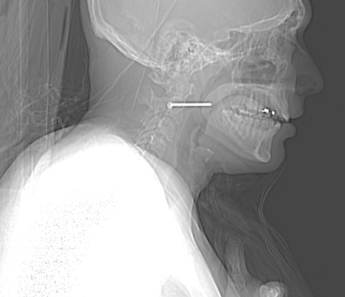
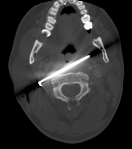
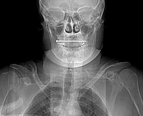
Here’s a post from my archive describing a different way to remove the foreign body. This is the technique I used, instead of the standard neck incision. The final incision was just a slight extension of the puncture wound, measuring only 1cm. I was able to grasp the head and pull it out without difficulty. The surprising thing to me was the amount of force I needed to apply to actually pull it out! No bleeding, no problems. The patient was observed for 24 hours and discharged home. He had no complications.

A Cool Way To Remove Embedded Foreign Bodies

Many of us have had the experience of digging into bloody tissue for long periods of time trying to locate the object, even with fluoroscopy. Well, there’s a better way of doing this.

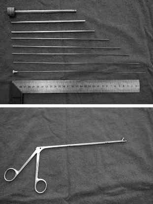
A group in China described a technique using a fancy form of needle localization. They employed a set of instruments normally used for lumbar diskectomy (see photo). This set includes a long 18 Ga needle with a removable hub, several dilators and an outer cannula with a 5.8mm diameter. A pair of 3.8mm grasping forceps is also used.

The foreign body is located using a C-arm fluoroscopy unit and the best approach is planned. The 18 Ga needle is then inserted using fluoro until it touches the object. The hub is removed and dilators are inserted over the needle, one after the other. The outer cannula is then placed over them, and the needle and dilators are then removed. The cannula is manipulated until the foreign body (or a part of it) is located within the cannula. It is then grasped and removed, along with the cannula if needed. If the object is too large to enter the cannula, the cannula is pulled back slightly and the grasper introduced past the end of it to grip and remove the foreign body.

The writers shared the details of 76 patients who had a total of 251 foreign bodies removed over a 6 year period. The depth varied from 2.5 to 8.5cm. Procedure time ranged from 8 to 15 minutes, and fluoro exposure varied from 1 to 4 minutes. Success rate was 100% (all foreign bodies were removed) and there were no complications.

Bottom line: This is a very slick technique that promises to dramatically increase the success rate and decrease complications from removing foreign bodies. The amount of time spent is much less than the brute force technique, as is the amount of soft tissue trauma. Large objects that cannot be grasped with these forceps cannot be removed with this method. Although I am a little concerned that the authors’ results were so perfect, it’s certainly worth a try!

Reference: Percutaneous extraction of deeply-embedded radiopaque foreign bodies using a less-invasive technique under image guidance. J Trauma 72(1):302-305, 2012.

Print Friendly, PDF & Email

Nail In The Neck: The Operation

We’ve made sure that our victim of the nail gun to the neck did not need an emergent operation. Vitals are stable, there’s no uncontrolled hemorrhage, and the patient is neurologically intact. We’ve imaged him using CT angiography, and the nail does not appear to have injured any vital structures.

How do we get it out of there? There are two things that need to be considered: where and how.

Rule of thumb: If a foreign body is located anywhere near vital structures, take it out in the OR, no matter how good you think the imaging is. It may be tempting to just pull it out in the ED, but resist! CT scans look so good, and they are so detailed, but they are not perfect. The ED does not have the equipment, personnel, or lighting necessary if something goes awry.

Rule of thumb: Use all information available to plan the removal procedure. In this case, the head of the nail is to the patient’s right. Therefore, it must be removed from the right side. The CT shows that the nail passes very close to the posterior pharynx, so it will need to be evaluated during the procedure.

This patient was taken to the operating room. During the intubation, direct laryngoscopy was carried out to carefully inspect the entire pharynx and larynx. No evidence of penetration was seen. The entire neck, face, and upper chest were prepped and draped (I like to go overboard in trauma cases; you never know what is going to happen). Fluoroscopy was available.

The classic operation would have been to make an incision along the sternocleidomastoid on the right side. The nail head would be approached directly. Since long, thin objects can be notoriously difficult to locate, fluoro can be very helpful. The exact position with respect to the carotid and jugular can be ascertained. Then the nail head is grasped with a clamp, and the nail gently pulled out along its axis. A nice, long wait for any evidence of bleeding should occur. The area can then be irrigated and the incision closed. Skin antibiotics can be given postop, but only one dose at most.

Having said that, I opted for a different approach based on an old blog post here. Tune in to the final post in this series to see what I really did.

Print Friendly, PDF & Email

Nail In The Neck: Part 2

This case involves an accidental nail gun injury to the neck. The patient is hemodynamically stable, neurologically intact, the airway is patent and not threatened, and there is no apparent hematoma. There is a small puncture near the sternocleidomastoid muscle on the right, fairly high on the neck. The nail is not palpable on either side. And the patient only complains of a little discomfort when he swallows.

What to do? First, the patient has passed all the initial decision points that would send us straight to the OR (ABC problems in ATLS jargon). But, per physical exam and initial imaging, the nail must obviously come out. We just have to figure out what we need to know before we take it out, and determine the best way to retrieve it.

Given the patient’s stability, additional imaging will be helpful. Views in different planes, and details of what the nail might have passed through will be invaluable. The recommended study is a CT angio of the neck. This will give good information about nearby structures and the vasculature. And software reconstructions will provide good 2D/3D information for removal planning. Here’s a lateral view.

image

The nail is located in front of the body of C2. It appears to be high enough to be near the pharynx, but well above trachea and esophagus. You can also see that the nail entered a little posteriorly, and travels right to left and forward.

Here’s a representative CT slice.

image

The nail enters behind the carotids (just above the bifurcation) and IJ on the right, and ends anterior to them on the left. It passes very close to the posterior pharynx. So neurovascular structures are intact, and the aerodigestive tract is a maybe (back of the pharynx).

Obviously, this thing has to come out. The question is, how to do it? For you surgeons out there, tell me your choice of approach, incision, and instrumentation. Tweet/X or leave comments! Answers in the next post.

Print Friendly, PDF & Email

What Would You Do? Nail In The Neck

Here’s a very interesting case for you. A construction worker was carrying an object inside a building WHILE HOLDING HIS NAIL GUN! As he passed through the door, his elbow hit the frame and he brushed his neck with the business end of the gun. Guess what happened?

He experienced sharp pain, then noted pain every time he swallowed. He checked himself out in the mirror, and there was a small puncture wound in the right side of his neck. He presented to his local ED, with the complaints just noted. He was hemodynamically stable and neurologically intact. His airway was patent, and he had minimal pain. The following image was obtained. The nail measures about 6cm in length.

Let me know (by X or comments) what additional information you think you need. I’ll discuss that in my next post. Then we’ll figure out what to do about this problem.

Print Friendly, PDF & Email

What’s With Those John / Jane Doe Names?

Oftentimes, trauma patients arrive, are not very responsive, and are not carrying an ID.  However, our electronic health record systems have a very difficult time with this. To expedite care, most hospitals developed a system of pseudo-names to be used until the patient can be identified.

Originally, these names were often “Doe” names. The classic ones were John Doe and Jane Doe. Obviously, more than two names were needed, so other first names were adopted to provide a sizable pool of pseudo-names.  Other disciplines, such as law enforcement, have used a similar system.

Where did the concept of John/Jane Doe come from? The origin is the British legal system, way back in the 18th century. In those days, landlords would start a “process of ejectment,” known these days as eviction, to rid their properties of landlords or deadbeat tenants. The legal process was a bit complex, so they would file for the process using a fake name to initiate it. They frequently selected John Doe or Richard Roe for their filings.

The reason the Doe and Roe names were used has been lost to history. There is speculation that these names were derived from certain deer species endemic to Britain. But honestly, no one knows for sure now.  Although this process was dropped in Great Britain in the mid-1800s, it persists in the US legal system. Remember the landmark legal case Roe v Wade?

The use of Doe names in electronic health records is rapidly fading as well. The list of name pairs ending with Doe is just not unique enough. There are too many opportunities to mix up similar names, especially if the last one is always the same. This can result in catastrophic errors if test results are misinterpreted, or a blood transfusion with incorrect ABO typing is given to the wrong patient.

Most trauma centers have adopted temporary naming systems consisting of two words or unique names. Some use number and letter sequences combined with another unique word. The real trick in the electronic medical record world is smoothly merging the records utilizing the pseudo-name with the patient’s previous records under their actual name.

The best practice for this varies by electronic record system and hospital. If done too early, the change may disrupt critical processes, such as the massive transfusion protocol. If done too late, it is difficult for trauma professionals to see any records the patient may have under their actual name. Each center must develop its own system for converting from John Doe to the real name.

Print Friendly, PDF & Email