The EMS Handoff: Opportunity for Improvement

Handoffs occur in trauma care all the time. EMS hands the patient off to the trauma team. ED physicians hand off to each other at end of shift. They also hand off patients to the inpatient trauma service. Residents on the trauma service hand off to other residents at the end of their call shift. Attending surgeons hand off to each other as they change service or a call night ends. The same process also occurs with many of the other disciplines involved in patient care as well.

Every one of these handoffs is a potential problem. Our business is incredibly complicated, and given that dozens of details on dozens of patients need to be passed on, the opportunity for error is always present. And the fact that resident work hours are becoming more and more limited increases the need for handoffs and the number of potential errors.

Today, I’ll look at information transfer at the first handoff point, EMS to trauma team. Some literature has suggested that there are 16 specific prehospital data points that affect patient outcome and must be included in the EMS report. How good are we at making sure this happens?

An observational study was carried out at a US Level I trauma center with video recording capabilities in the resuscitation room. Video was reviewed to document the “transmission” part of the EMS report. Trauma chart documentation was also reviewed to see if the “reception” half of the process by the trauma team occurred as well.

Here are the factoids:

  • A total of 96 handoffs were reviewed over a one year period
  • The maximum number of data elements in the study was 1536 (96 patients x 16 data elements)
  • The total number “transmitted” was 473, but only 329 of those were “received.”
  • This is not quite as bad as it seems, since 483 points were judged as not applicable by the reviewers. However, this left 580 that were applicable but were not mentioned by EMS.
  • Of the 16 key elements, the median number transmitted was 5, with a range of 1-9.

This sounds bad. However, the EMS professionals and the physicians have somewhat different objectives. EMS desperately wants to share what they know about the scene and the patient. The trauma team wants to start the evaluation process using their own eyes and hands. What to do?

Bottom line: EMS to trauma team handoffs are a problem for many hospitals. EMS has a lot of valuable information, and the trauma team wants to keep the patient alive. They are both immersed in their own world, working to do what they think is best for the patient. Unfortunately, they could do better if the just worked together a bit more.

Tomorrow I’ll share a solution to the EMS-trauma team handoff problem.

Reference: Information loss in emergency medical services handover of trauma patients. Prehosp Emerg Care 13:280-285, 2009.

Does Trauma Team Prenotification By EMS Decrease Mortality?

A few months ago, I heard this statement at a conference I was attending:

“Of course, prenotification of the trauma team by EMS decreases hospital mortality”

And of course, whenever I hear someone say “of course”, it makes me think about it. How do we know for sure? So I made one of my frequent trips to PubMed to find the basis for the statement.

And guess what? He shouldn’t have said “of course.” The literature is very scarce on this topic. There are actually some good papers detailing the advantages of prehospital notification for things like stroke and STEMI. But trauma?

A group in Melbourne, Australia performed a systematic review of the literature on this topic for the Australia-India Trauma System Collaboration. They were interesting in finding information about early (<24 hour) and overall (<30 day) mortality, as well as trauma team presence, time to critical hospital interventions, and hospital length of stay. Over a thousand articles were identified, but half did not have proper study design, and a quarter weren’t about notification. After excluding those, and others that failed other criteria, they were left with only three to review!

Here are the factoids:

  • Two of the studies were small, with only 81 and 269 participants and individual hospitals
  • The remaining study was a very large retrospective analysis of over 72,000 patients from 59 hospitals in Canada
  • All three had serious risk for bias and significant confounding variables
  • The large study showed a significant improvement in overall mortality from 32% to 23%, the smaller studies did not. But the study quality was so poor for this outcome that we can’t really be certain, and these numbers seem very high coming from Canada.
  • No conclusions could be drawn for short term mortality, length of stay, or time to interventions in the ED
  • The studies only involved high-income countries; nothing could be learned for low to medium-income countries.

Bottom line: Three studies in 27 years??! So sad. It certainly seems like having the trauma team informed and prepped in advance should count for something. But like so many other things in this business, we just don’t know for sure. Having everyone in place and ready to receive the patient, and getting other in-hospital resources ready (e.g. OR) may shorten time to definitive, life-saving treatment. But for now, we’ll just have to pretend. Until someone designs and performs a much better study.

Related posts:

Reference: Prehospital notification for major trauma patients requiring emergency hospital transport: A systematic review. J Evidence Based Med 10(3):212-221, 2017.

Chest X-Ray After Chest Tube Insertion: Why Do We Do It?

More dogma, or is it actually useful? Any time a chest tube (tube thoracostomy) is inserted, we automatically order a chest x-ray. Even the ATLS course recommends obtaining an image after placement. But anything we do “automatically” is grounds for critical analysis to see if there is a valid reason for doing it.

A South African group looked at the utility of this practice retrospectively in 1004 of their patients. They place 1042 tubes. Here are the factoids:

  • Patients were included if they had at least one chest x-ray obtained after insertion
  • Patients were grouped as follows: Group A (10%) had the tube inserted on clinical grounds with no pre-insertion x-ray (e.g. tension pneumothorax). Group B (19%) had a chest x-ray before and had ongoing clinical concerns after insertion. Group C (71%) had a chest-xray before and no ongoing concerns.
  • 75% of injuries were penetrating (75% stab, 25% GSW), 25% were blunt
  • Group A (insertion with pre-x-ray): 9% had post-insertion findings that prompted a management change (kinked, not inserted far enough)
  • Group B (ongoing clinical concerns): 58% required a management change based on the post-x-ray. 33% were subcutaneous or not inserted far enough (!!)
  • Group C (no ongoing clinical concerns): 32 of 710 (5%) required a management change, usually because the tube was too deep

The authors concluded that if there are no clinical concerns (tube functioning, no clinical symptoms) after insertion, then a chest x-ray is not necessary.

Bottom line: But I disagree with the authors! Even with no obvious clinical concerns, the tube may not be functioning for a variety of reasons. Hopefully, this fact would then be discovered the next day when another x-ray is obtained. But this delays the usual progression toward removing the tube promptly by at least one day. It increases hospital stay, as well as the likelihood of infection or other hospital-associated complication. A chest x-ray is cheap compared to a day in the hospital, which would potentially happen in 5% of these patients. I recommend that we continue to obtain a simple one-view chest x-ray after tube insertion.

Related posts:

Reference: What is the yield of routine chest radiography following tube thoracostomy for trauma?  Injury 46(1):45-48, 2015.

Is Applying Or Removing That Cervical Collar Dangerous?

Cervical collars are applied to blunt trauma patients all the time. Maybe a bit too often. And most of the time, the neck is fine. It’s just those few patients that have fracture or ligamentous injury that really, truly need it.

I’ve previously written about how good some of the various types of immobilization are at limiting movement (click here). But what happens when you are actually putting them on or taking them off? Could there be dangerous amounts of movement then?

Several orthopaedics departments studied this issue using an electromagnetic motion detector on “fresh, lightly embalmed cadavers” (!) to determine how much movement occurred when applying and removing 1- and 2-piece collars. Specifically, they used an Aspen 2-piece collar, and an Ambu 1-piece. They were able to measure flexion/extension, rotation and lateral bending.

Here are the factoids:

  • There were no significant differences in rotation (2 degrees) and lateral bending (3 degrees) when applying either collar type or removing them (both about 1 degree)
  • There was a significant difference (of 0.8 degrees) in flexion/extension between the two types (2-piece flexed more). Really? 0.8 degrees is significant? Not clinically!
  • Movement was similarly small and not significantly different in either collar when removing them

Bottom line: Movement in any plane is less than 3-4 degrees with either a 1-piece or 2-piece collar. This is probably not clinically significant at all. Just look at my related post below, which showed that once your patient is in the rigid collar, they can still flex (8 degrees), rotate (2 degrees) and move laterally (18 degrees) quite a bit! So be careful when using any collar, but don’t worry about doing damage if you use it correctly.

Related post:

Reference: Motion generated in the unstable cervical spine during the application and removal of cervical immobilization collars. J Trauma 72(6):1609-1613, 2012.

Sinus Precautions: Another Trauma Urban Legend?

The trauma service typically helps our facial surgery colleagues manage patients from time to time. Every now and again, I see their request/instructions for “sinus precautions” when patients have certain fracture patterns or undergo surgery involving sinuses.  Where did these “precautions” come from? Do they really make sense?

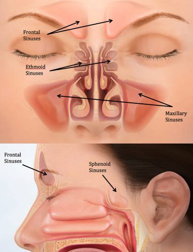
The paranasal sinuses shown above are the most commonly injured after facial trauma. They are lined with mucosa, and like the rest of our body, colonized with bacteria. They are interconnected with each other and the nasal cavity via tiny ostia that allow for normal drainage into the nose. Facial surgeons worry that these bacteria may seep out of fractured areas into clean tissue and cause deep infections. This may occur spontaneously, and could be accelerated if the pressure in the sinuses is increased in any way.

If you do a simple internet search on “sinus precautions”, it seems like every facial surgery group in the country has a set in their patient information sheet. And they all look suspiciously similar. As if they’ve been copied from each other and over, and from the same very, very old document.

As noted above, the main issue to be avoided is increasing pressure across the sinuses. Here’s the usual list of precautions, and my comments on their utility.

  • Avoid blowing your nose. This one actually makes sense. Closing the nose and mouth, pressurizing the pharynx and releasing through the nose will certainly increase sinus pressures. And there are actually reported cases of new onset cellulitis after this maneuver. True
  • Avoid sneezing. Good luck on this one. Who actually does this on purpose anyway? Sneezing will certainly increase sinus pressures, but this can be minimized by avoiding trying to stifle the sneeze. True
  • Do not drink through a straw. What? This causes a low pressure zone in the oropharynx so that fluids can be sipped, but the nasopharynx is isolated. If it weren’t you’d have beverages pouring out of your nose. False
  • Do not smoke. Well, this is certainly a good idea for many reasons, but has little impact on nasopharyngeal pressures. If anything, it decreases them slightly on inhalation, and works the same on exhalation as breathing out without the smoke. False
  • Do not blow musical instruments, balloons, … Yes, this can increase pressures. True
  • Do not push or lift heavy objects.  Hmm. The natural tendency is to perform a Valsalva maneuver (holding breath against a closed glottis) when doing this. In theory, this shouldn’t impact the sinuses because they are not attached to the trachea in any way. And even though it is possible to let some air escape the glottis and keep your mouth closed, it immediately exits the nose. Exhaling through the nose rapidly could increase pressures slightly. Most people don’t do this. Mostly false
  • Do not bend over, keep your head above your heart, sleep with your head slightly elevated. Come on, now. Sinuses are rigid, air-filled cavities. They don’t compress. False
  • Do not fly in a plane.  Definitely an issue. Everybody experiences popping ears and sinuses when flying. And it’s the descent that is of most concern. The increasing air pressure during this phase of flight can push fluid and air out of the sinuses. True
  • Do not spit. Okay, this goes without saying. It’s rude, but doesn’t do a thing to your sinuses. False

Bottom line: I was unable to find any seminal paper or book chapter as the source for “sinus precautions”. Most of the items on the list are bogus. But a few do actually increase sinus pressures and could result in fluid, air, and bacteria moving out of sinuses and into areas where they don’t belong. Pare down the list before you hand it to your patients.