All posts by The Trauma Pro

First, There Was REBOA, And Now… GROA?!

REBOA (Resuscitative Endovascular Balloon Occlusion of the Aorta) is a “newer” resuscitative technique that has actually been around since the Korean War. It was first used to treat two injured soldiers, and although they ultimately died, it spurred research into the technique and its applications.

Balloon occlusion techniques were then adopted by vascular surgeons and were found to be useful as an adjunct in managing ruptured abdominal aneurysms. A slow trickle of studies on human use in trauma began to surface. But when autopsy studies carried out during the Gulf War showed that uncontrolled torso hemorrhage was a major cause of death, research in the technique exploded.

First, there were a rash of pig studies evaluating the feasibility of using a percutaneously placed occlusion device in the early 2010s. This transitioned to human studies around 2014, and after that we were off to the races. Over 100 papers on REBOA are now published each year.

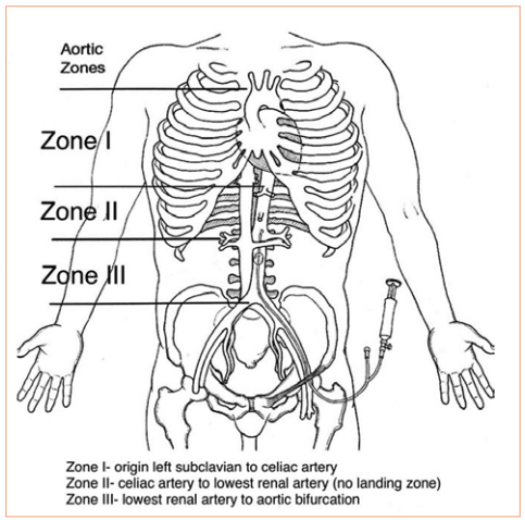
REBOA has been shown to have some advantage in patients with abdominal or pelvic sources of bleeding. The catheter is inserted in the groin and the balloon inflated in one of two zones, depending on the location of the hemorrhage (see diagram below). For abdominal bleeding, it is inserted just above the diaphragm in Zone I. For pelvic bleeding, it is inserted below the takeoff of the visceral arteries and above the aortic bifurcation, in Zone III.
In the US, REBOA catheters are only inserted upon arrival to the hospital. There are a few random reports of field placement where a physician is part of the prehospital team. By definition, this technique is generally not available in austere environments, only upon arrival in the emergency department.

Researchers at the University of Michigan began looking for an alternative technique that could be applied in the field by non-physicians. They noted the close anatomic relationship of the distal esophagus, proximal stomach, aorta, and thoracic vertebrae, and designed a device to compress the aorta against the spine in this area.

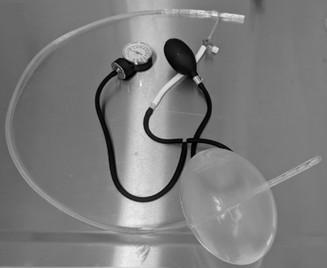
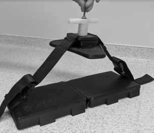
They developed a prototype device which they named GROA (gastro-esophageal resuscitative occlusion of the aorta). It consists of a gastro-esophageal tube with an ovoid balloon, an air pump with pressure measurement device, and an external compression device. Here is a picture of the device:
And here’s a diagram of what it looks like when inserted:
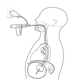
The tube is inserted and the balloon inflated. The external compression device is then placed around the patient, with the top plate located over the epigastrium and the bottom plate under the patient. It is designed to apply anterior pressure over the balloon, but to avoid circumferential constriction of the abdomen.
Bottom line: This device is an interesting development in the balloon occlusion space. As with early studies of REBOA, GROA is currently being investigated using a pig model. If it appears to be beneficial, it will still be several years before it makes the jump to human subjects. If effective, this concept would allow prehospital providers to apply some degree of hemorrhage control when it originates in the abdominal cavity.

There are currently exactly three papers on this new technique, and I have included the references below if you are interested in reading them. I’m sure there are many more to come and it may eventually be competing with REBOA for journal space.

There is one consideration to be aware of when reading these papers that is similar to much of the research on REBOA. Two of the authors have a financial interest in the company that licenses the GROA technology. And in the most recent study, another one of the authors is an advisory board member for one of the manufacturers of REBOA catheters. For these reasons, it is less likely that they will publish papers that are not favorable to the product. So read critically!

References:

  • Gastroesophageal resuscitative occlusion of the aorta: Physiologic tolerance in a swine model of hemorrhagic shock, Journal of Trauma and Acute Care Surgery: December 2020 – Volume 89 – Issue 6 – p 1114-1123 doi: 10.1097/TA.0000000000002867
  • Gastroesophageal resuscitative occlusion of the aorta prolongs survival in a lethal liver laceration model, Journal of Trauma and Acute Care Surgery: May 2022 – Volume 92 – Issue 5 – p 880-889 doi: 10.1097/TA.0000000000003444
  • Tandem use of Gastroesophageal Resuscitative Occlusion of the Aorta followed by REBOA in a Lethal Liver Laceration Model, Journal of Trauma and Acute Care Surgery: June 10, 2022 –  doi: 10.1097/TA.0000000000003719

MRI And External Fixators

MRI is an indispensable tool for evaluation of spine and soft tissue trauma. However, a great deal of effort was be made to ensure that any patient scheduled for this test is “MRI compatible.” The fear is that any retained metallic fragments may move or heat up once the magnets are activated.

But what about trauma patients with external fixators? That is one big hunk of metal that is inserted deep into your patient. There are three major concerns:

  • Is the material ferromagnetic? If so, it will move when the magnets are activated and may cause internal injury. These days, there are many fixator sets that are not ferromagnetic, avoiding this problem.
  • Can currents be induced in the material, causing heating? This is not much of a problem for small, isolated objects. However, external fixators are configured in such a way that loops are created. The fluctuating magnetic fields can induce currents that in turn will heat the surrounding tissue. And thinner materials (narrow pins) result in more current and more heating.
  • Will the metal degrade image quality?

The biggest challenge is that there is no standard ex-fix configuration. Our orthopaedic colleagues get to unleash their creativity trying to devise the appropriate architecture to hold bones together so they can heal properly. This makes it difficult to develop standardized guidelines regarding what can and can’t go into the scanner.

However, there is a growing body of literature showing that the heating effects are relatively small, and get smaller as the distance from the magnet increases. And non-ferromagnetic materials move very little, if at all, and do not interfere with the image. So as long as nonferromagnetic materials are used, the patients are probably safe as long as basic principles are adhered to:

  • Other diagnostic options should be exhausted prior to using MRI.
  • Informed consent must be obtained, explaining that the potential risks are not completely understood.
  • The fixator must be tested with a handheld magnet so that all ferromagnetic components can be identified and removed.
  • All traction bows must be removed.
  • Ice bags are placed at all skin-pin interfaces.
  • The external fixator must remain at least 7cm outside the bore at all times.

Bottom line: MRI of patients with external fixators can be safely accomplished. Consult your radiologists and physicists to develop a policy that is specific to the scanners used at your hospital. 

Related posts:

Chest Tube Based On Pneumothorax Size

How big is too big? That has been the question for a long time as it applies to pneumothorax and chest tubes. For many, it is a math problem that takes into account the appearance on chest x-ray, the physiology of the patient, and their ability to tolerate the pneumothorax based on any pre-existing medical conditions.

I first wrote about this paper when it was just an abstract for last year’s AAST meeting. Apparently, it passed peer review muster. It has just been published in the Journal of Trauma. The numbers have changed a little bit, so I’ll update my analysis accordingly.

The group at Froedtert in Milwaukee has been trying to make the decision to place a chest tube a bit more objective. They introduced the concept of CT based size measurement using a 35mm threshold at the AAST meeting three years ago. Read my review here. My criticisms at the time centered around the need to get a CT scan for diagnosis and their subjective definition of a failure requiring chest tube insertion. The abstract never did make it to publication.

The authors are back now with a follow-on study. This time, they made a rule that any pneumothorax less than 35mm from the chest wall would be observed without tube placement. The performed a retrospective review of their experience and divided it into two time periods: 2015-2016, before the new rule, and 2018-2019, after the new rule. They excluded any chest tubes inserted before the scan was performed, those that included a sizable hemothorax, and patients placed on a ventilator or who died.

Here are the factoids:

  • There were 99 patients in the early period and 167 in the later period
  • Chest tube use significantly declined from 28% to 18% between the two periods. These numbers are 8% higher than were described in last year’s abstract.
  • Observation rates without a chest tube increased from 85% to 95% after implementation of the new guideline
  • There was no difference in length of stay, inpatient failure rate, complications, or death
  • The most common inpatient failure was due to development of a new hemothorax. However, there was an almost identical number of failures of “unclear” etiology. This is troublesome but part and parcel for such a retrospective study.
  • Two patients were readmitted within 30 days for a pulmonary complication (one empyema, one readmission at 3 days after discharge for dyspnea due to pneumothorax)
  • Patients in the later group were 2x more likely to be observed (by regression analysis)

The authors concluded that the 35mm rule decreased unnecessary chest tube insertion while maintaining patient safety.


Bottom line: I still have a few issues with this paper and the authors’ preceding series of abstracts. First, decision to insert a chest tube required a CT scan in a patient with a pneumothorax. This seems like extra radiation for patients who may not otherwise fit any of the usual blunt imaging criteria. And, like their 2018 and 2021 abstracts, there are no objective criteria for failure requiring tube insertion. So it is difficult to gauge compliance when insertion for failure is somewhat based on the whims of the individual surgeon.

What this abstract really shows is that compliance with the new rule increased, and there were no obvious complications from its use. The other numbers (chest tube insertions, observation failure) are just too subjective to learn much from. The most troubling issue is that the reason for 40% of failures was “unclear.” This is most likely due to the fact that the authors did not have objective guidelines for failure due to the retrospective nature of the study.

The numbers in this paper changed a little from last year’s abstract. The overall conclusions and meaning did not. It appears that 35mm is a reasonable threshold for pneumothorax size when contemplating inserting a chest tube. Unfortunately, this study relied entirely on CT scan. We don’t know if using a similar guideline for regular old chest x-ray is valid or not. 

What we still need is a good, prospective trial using an arbitrary guideline like 35mm pneumothorax as seen on chest x-ray or CT scan. And then, a clear definition of what defines a failure that requires tube insertion would be helpful. And at some point, we also need to know if a small tube or pigtail catheter is adequate for pure pneumothorax. Don’t get me started on that one!

Reference: The 35-mm rule to guide pneumothorax management: Increases appropriate observation and decreases unnecessary chest tubes. J Trauma 92(6):951-957, 2022.

You’ve Been Pimped! Origins And How To Survive It!

What exactly is pimping? If you have ever been a medical student or resident in any discipline, you probably already know. It’s ostensibly a form of Socratic teaching in which an attending physician poses a (more or less) poignant question to one or more learners. The learners are then queried (often in order of their status on the seniority “totem pole”) until someone finally gets the answer. But typically, it doesn’t stop there. Frequently, the questioning progresses to the point that only the attending knows the answer.

So how did this time honored tradition in medical education come about? The first reference in the literature attributes it to none other than William Harvey, who first described the circulatory system in detail. He was disappointed with his students’ apparent lack of interest in learning about his area of expertise. He was quoted as saying “they know nothing of Natural Philosophy, these pin-heads. Drunkards, sloths, their bellies filled with Mead and Ale. O that I might see them pimped!”

Other famous physicians participated in this as well. Robert Koch, the founder of modern bacteriology, actually recorded a series of “pümpfrage” or “pimp questions” that he used on rounds. And in 1916, a visitor at Johns Hopkins noted that he “rounded with Osler today. Riddles house officers with questions. Like a Gatling gun. Welch says students call it ‘pimping.’ Delightful.”

So it’s been around a long time. And yes, it has some problems. It promotes hierarchy, because the attending almost always starts questions at the bottom of the food chain. So the trainees come to know their standing in the eyes of the attending. And they also can appreciate where their fund of (useful?) knowledge compares to their “peers.” It demands quick thinking, and can certainly create stress. And a survey published last year showed that 50% of respondents were publicly embarrassed during their clinical rotations. What portion of this might have been due to pimping was not clear.

Does pimping work? Only a few small studies have been done. Most medical students have been involved with and embarrassed by it. But they also responded that they appreciated it as a way to learn. A 2011 study compared pimping (Socratic) methods to slide presentations in radiology education. Interestingly, 93% preferred pimping, stating that they felt their knowledge base improved more when they were actively questioned, regardless of whether they knew the answer.

So here are a few guidelines that will help make this technique a positive experience for all:

For the “pimpers”:

  • Make sure that the difficulty level of questions is reasonable. You are testing your learners’ knowledge, not spotlighting your own mental encyclopedia
  • Build the level of difficulty from questions that most can answer to one or two that no one knows, then switch to didactice teaching of the esoterica
  • Don’t let one learner dominate the answers; gently exclude them and solicit answers from others so they get a chance to participate
  • Provide positive reinforcement for correct answers, but don’t resort to negative reinforcement (insults) when they are wrong
  • Go Socratic when the answer is not known. Step back and review the basic concepts involved that helps your learners arrive at the correct answer.

For the “pimpees”:

  • Read, read, read! You are in this to learn, so study all the clinical material around you.
  • Talk to your seniors to find out your attending’s areas of interest. There’s a lot of stuff to learn, and this may help you focus your rounding preparation a bit. It still doesn’t absolve you from learning about all the other stuff, though.
  • Don’t be “that guy (or gal)” who tries to dominate and answer every question
  • If all else fails, and it’s one of those “percentage” questions, use my
    “85/15 rule.”
    If the issue you are being asked about seems pretty likely, answer “85%.” If it seems unlikely, go with “15%.” It’s usually close enough to the real answer to satisfy.

Bottom line: Pimping is a time-honored tradition in medicine, but should not be considered a rite of passage. There is a real difference in attitudes and learning if carried out properly. Even attendings have a thing or two to learn about this!

Reference: The art of pimping. JAMA. 262(1):89-90, 1989.

You’ve Been Pimped!

You know what I’m talking about. It’s a mainstay of medical education for physicians. It starts in medical school, and generally never stops. And when you finish your residency,  you graduate from being pimped to being the pimper.

How did this all come to be? Is it good for education? Bad? Tune in tomorrow to learn more. In the meantime, enjoy this algorithm on how to get through a pimping session. Click to view full-size.

pimping

Source: Posted by Dr. Fizzy on The Almost Doctor’s Channel