All posts by The Trauma Pro

Part 1: Metal Splints – Can You X-ray Through Them?

Splinting is an important part of the trauma resuscitation process. No patient should leave your trauma resuscitation room without splinting of all major fractures. It reduces pain, bleeding, and soft tissue injury, and can keep a closed fracture from becoming an open one.

But what about imaging? Can’t the splint degrade x-rays and hamper interpretation of the fracture images? Especially those pre-formed aluminum ones with the holes in them? It’s metal, after all.
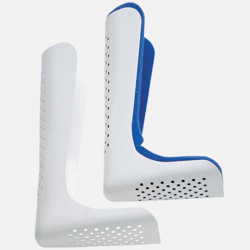
Some of my orthopedic colleagues insist that the splint be removed in the x-ray department before obtaining images. And who ends up doing it? The poor radiographic tech, who has no training in fracture immobilization and can’t provide additional pain control on their own.

But does it really make a difference? Judge for yourself. Here are some knee images with one of these splints on:
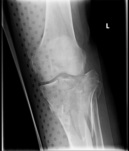
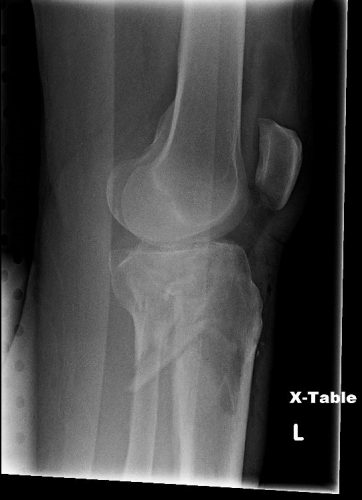
Amazingly, this thin aluminum shows up only faintly. There is minimal impact on interpretation of the tibial plateau. And on the lateral view, the splint is well posterior to bones.
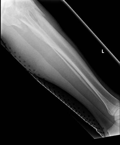
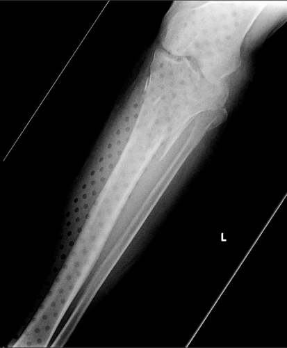
On the tib-fib above, the holes are a little distracting on the AP view, but still allow for good images to be obtained.

Bottom line: In general, splints should not be removed during the imaging process for acute trauma. For most fractures, the images obtained are more than adequate to define the injury and formulate a treatment plan. If the fracture pattern is complex, it may be helpful to temporarily remove it, but this should only be done by a physician who can ensure the fracture site is handled properly. In some cases, CT scan may be more helpful and does not require splint removal. And in all cases, the splint should also be replaced immediately at the end of the study.

In my next post, I’ll look at the use of CT scans when this type of splint is in use.


How To: Insert A Small Percutaneous Chest Tube

This short (10 minute) video demonstrated the technique for inserting small chest tubes, also known as “pigtail catheters.” It features Jessie Nelson MD from the Regions Hospital Department of Emergency Medicine. It was first shown at the third annual Trauma Education: The Next Education conference in September 2015, for which she was a course director.

Please feel free to leave any comments or ask any questions that you may have.

YouTube player

Related posts:
Pigtail catheters vs regular chest tubes
Tips for regular chest tubes 

A Blood Test For TBI? Part 3

The FDA announced approval of a blood test that incorporates both GFAP and UCH-L1. Approval was based on two as yet to be published studeis titled Evaluation of Biomarkers of Traumatic Brain Injury (ALERT-TBI) and Evaluation of Biomarkers of Traumatic Brain Injury Extension Study (ALERT-TBIx), and passed after less than 6 months of evaluation. Yes, more silly acronyms, I know.

The studies were designed to “evaluate the utility of the Banyan UCH-L1/GFAP Detection Assay as an aid in the evaluation of suspected traumatic brain injury (Glasgow Coma Scale score 9-15) in conjunction with other clinical information within 12 hours of injury to assist in determining the need for a CT scan of the head.”

The former study started in 2012 and involved 2011 patients! The latter had only 119 patients, starting in 2015. Now, I have no access to their data, so I can’t tell what the FDA saw.

From Banyan Biomarkers’ website:

“The CT scan is widely available to assist clinicians in the evaluation of TBI, however, CT scans do not provide a clear and objective answer and scanning may increase the risk for radiation-induced cancer. Furthermore, over 90% of patients presenting to the emergency department with mild TBI, sometimes described as “concussion”, have a negative CT scan. Despite these limitations, nearly all patients are sent for a CT, which results in increased costs to the healthcare system and unnecessary patient exposure to radiation.”

Here are the (very) few factoids that I can find:

  • CT scan results were compared to the Brain Trauma Indicator (BTI) blood test (GFAP + UCH-L1)
  • BTI predicted a positive CT scan 98% of the time
  • It predicted a negative CT scan 99.6% of the time
  • Time to process the test is currently 4 hours

Bottom line: Sounds promising, right? Based on the data summarized over the last two days, I wouldn’t be too excited about this test, but the FDA was able to look at a study that I can’t. It appears that the negative predictive value is excellent, so I can see the application.

That being said, 4 hours is way to long. We can’t have a patient sitting in the ED waiting for the results to come back to decide whether they need a head CT. And how long will it take the assay to be widely available?

The devil will be in the details. What types of intracranial lesions were detected. Are the negative predictive values the same for subarachnoid, subdural, epidural, or intraparenchymal bleeds? And finally, how expensive will it be? How does the cost for the test compare to the cost of a CT scan done in 5 minutes?

I’ll let you know more as the details emerge. But don’t look for, or plan to use, this test at your hospital any time soon. There’s more work to do!

Reference: Banyan Biomarkers (banyanbio.com)

A Blood Test For TBI? Part 2

Yesterday, I wrote about one blood biomarker, GFAP, and its possible application in detecting traumatic brain injury (TBI). Today, I’ll discuss a complementary marker called UCH-L1.

Fewer studies have been done looking at the utility of UCH-L1 in detecting TBI than of GFAP.  A review article published last month pooled existing literature to get a sense of how good this biomarker really is. It also examined the risk of bias due to the small numbers of studies involved.

Here are the factoids:

  • Only 38 abstracts were eligible, but full text was available for analysis in only 13 (meaning it was only an abstract and never passed muster for publication). The authors of the published studies were contacted for additional information, which is an interesting (and helpful) practice.
  • Of all of those, only 4 were selected for meta-analysis! This significantly limited the value of the analysis.
  • Serum UCH-L1 has a high accuracy in predicting CT findings in mild to moderate TBI, but there is a high risk of bias affecting this result
  • Plasma UCH-L1 has a moderate accuracy predicting CT findings across all GCS levels, with a low risk of bias
  • Pooling all studies, this is high accuracy in predicting CT findings in patients with TBI across all GCS levels, but there is a high risk of bias affecting the results

Bottom line: UCH-L1 show promise as a predictor of CT findings in patients with TBI. However, the research papers were few and far between, and the possibility of bias was high. What does this mean? That using this test alone is better than a coin toss, but not good enough to change our practice in ordering CT scans in head injured patients. More well-designed studies are needed tell us whether this new (and undoubtedly expensive) test is worth the trouble.

Tomorrow, I’ll discuss a blood test incorporating both UCH-L1 and GFAP that was recently approved by the FDA.

Reference: The diagnostic values of UCH-L1 in traumatic brain injury: A meta-analysis. Brain Inj 32(1):1-17, 2018.