Tag Archives: VTE

Routine Duplex Screening For Venous Thromboembolism

Venous thromboembolism (VTE) is a potential problem for all hospitalized patients, and traumatic injury is yet an additional risk factor for its occurrence. Most trauma centers have some kind of risk assessment tool to help the tailor their chemoprophylaxis regimen to patients most at risk. But far fewer have adopted the use of screening ultrasounds to monitor for new onset VTE that would dictate conversion to therapeutic treatment.

Unfortunately, in the US, the Centers for Medicare and Medicaid Services (CMS) has deemed VTE as a “never” event and penalizes hospitals when they report it. One of the unintentional consequences of this (or is it?) is that hospitals may then pressure trauma programs to avoid surveillance in order to “make the numbers look better.” Remember Law X from Samuel Shem’s House of God?

X. If you don’t take a temperature, you can’t find a fever.

Similarly, if you don’t do a duplex screen, you probably won’t detect VTE. Now granted. some patients develop classic symptoms like edema, pain, and tenderness. But not that many.

But is this wise? My contention has been that if the patient doesn’t develop symptoms that catch your attention, yet they develop VTE that you don’t know about, they are at risk for more serious complications like pulmonary embolism (PE). And you are blithely unaware.

The trauma group at Intermountain Medical Center in Salt Lake City performed an elegant study to determine the impact of screening for VTE in their trauma patients. They performed a prospective, randomized trial on trauma patients admitted over a 30-month period. Patients were included if they were judged to be at moderate to high risk based on their risk assessment profile (RAP) score. Patients were excluded if they were children, had VTE or PE within 6 months prior to hospitalization, or had some type of hypercoagulable state.

Patients were sequentially randomized to no duplex screening vs screening on days 1, 3, 7, and then weekly thereafter. The primary outcome measure was PE during the hospital stay. Secondary outcomes consisted of a number of factors relating to development of DVT.

Here are the factoids:

  • Nearly two thousand patients were enrolled, with about 995 patients in each group and no differences in demographics
  • The ultrasound group had significantly more below-knee (124 vs 8) and above knee (19 vs 8) DVT identified (no surprise there)
  • The ultrasound group had significantly fewer pulmonary emboli than the no ultrasound group (1 vs 9) (lots of surprise here!)
  • Mortality was similar during the hospital stay and for 90 days after

Bottom line: If you look for it, you will find it! This is the definition of surveillance bias. But in in this study, looking for clots in the legs may also decrease the number of patients who develop symptomatic pulmonary embolism. How could this be?

There are a few possibilities. The majority of DVT found in the surveillance group were located distally. Although there is some uncertainty as to how likely these are to embolize, it is probably very low. So let’s ignore them for now and assume that only the proximal clots might embolize.

This leaves an extra 11 DVT found in the surveillance group over and above the no-ultrasound group. Despite that, the surveillance group had only one PE vs 9 in the no-ultrasound group!

Another explanation was that the ultrasound guided changes in management, shifting to management to therapeutic drug dosing. The authors did not find a significant difference between the use of therapeutic vs prophylactic dosing between the groups. But there was a difference. Although the overall study was well-powered, there really weren’t enough numbers to show whether there was a true difference in therapeutic dosing. Fourteen patients in the ultrasound group got therapeutic anticoagulation compared to only 4 in the no-surveillance group. I think this is the actual reason.

Overall, this is a well-designed and well-executed study that shows why taking the Ron Popeil approach to DVT prophylaxis (“set it and forget it”) doesn’t work. Patients do occasionally develop proximal DVT on standard chemoprophylaxis (and frequently develop distal DVT), but it doesn’t always result in obvious signs and symptoms. This study shows that if you don’t look for it, you may not know until they suddenly develop chest pain, air hunger, and worse! So consider carefully if your practice guideline doesn’t yet include surveillance.

Reference: Trauma Patients at Risk for Venous Thromboembolism who Undergo Routine Duplex Ultrasound Screening Experience Fewer Pulmonary Emboli: A Prospective Randomized Trial. J Trauma, publish ahead of print, Publish Ahead of Print. DOI: 10.1097/TA.0000000000003104, February 4, 2021.

Should I Apply Compression Devices To Patients With DVT?

Everyone knows that venous thromboembolism (VTE) is a potential problem in hospitalized patients, and especially so in trauma patients. Several groups of them are at higher risk by virtue of the particular injuries they have sustained and the activity restriction caused.

Nearly every trauma program uses some form of screening and prophylaxis in an attempt to reduce the occurrence of this problem, which can result in deep venous thrombosis (DVT) and/or pulmonary embolism (PE). Screening looks at patient factors such as age, obesity, previous VTE as well as injury risk factors like spine and pelvic fractures, and decreased mobility.

Based on the screening protocol, prophylaxis may be prescribed depending upon level of VTE risk, which is then balanced with bleeding risk from brain, solid organ, or other injuries. The choices we have are primarily mechanical vs chemical and consist of compression devices (sequential or not) and various heparins.

An age old question surfaced on my own patient rounds recently. If a patient breaks through their prophylaxis and develops DVT, is it safe to apply compression devices to the extremity?

There has always been the fear that doing things that increase flow in the affected extremity may cause clots to dislodge and ultimately cause a PE. Seems logical right? But we know that often, our common sense about things is completely wrong.  Couldn’t just moving around cause pieces to break off? A meta-analysis of 13 studies published in 2015 showed that early ambulation was not associated with a higher incidence of new PE. Furthermore, patients who suffered from pain in the affected extremity noted significant improvements with early ambulation.

If ambulation makes the pain better, could the veins be recanalizing more quickly? Another study examined a small group of 72 people with DVT receiving anticoagulants, half of whom were prescribed exercise and compression stockings and the other half stockings only. There was a huge amount of variability in the rates of recanalization, but ultimately there were no significant differences with or without exercise.

So just lying in bed is not good, and exercise/ambulation may actually make people feel better. But interestingly, bedrest alone does not appear to increase the likelihood of PE! It does decrease the risk of developing problems other than the VTE, like pulmonary complications.

But what about compression devices? Common sense would say that you are intermittently  increasing pressures in the leg veins, which could dislodge any loose clots and send them flying to the lungs, right?

Unfortunately, I couldn’t find a paper from anyone who had the courage to try this. Or perhaps no institutional review board (IRB) would approve it. But the key fact is that every compression device manufacturer includes existing DVT as a contraindication in their product documentation. They don’t have any literature either, so I assume it’s an attempt to limit litigation, just in case.

Bottom line: Walking provides at least as much muscle compression as compression devices. But the simple truth is that we have no solid research that either supports or condemns the use of active compression devices in patients with known DVT. And we probably won’t, ever.

Compression stockings seem to be safe, but they really don’t do much. They are white, but don’t do much more than contribute to hospital clothing fashion. Since the manufacturers define existing DVT as a contraindication, application of their product would be considered an off-label use. So it looks like we cannot in good faith use these devices in patients with diagnosed DVT.

References:

  • Bed Rest versus Early Ambulation with Standard Anticoagulation in The Management of Deep Vein Thrombosis: A Meta-Analysis. PLOS One , April 10, 2015, https://doi.org/10.1371/journal.pone.0121388
  • Bed Rest or Ambulation in the Initial Treatment of Patients With Acute Deep Vein Thrombosis or Pulmonary Embolism: Findings From the RIETE Registry. Chest 127(5):1631-1636, 2005.
  • Does supervised exercise after deep venous thrombosis improve recanalization of occluded vein segments? A randomized study. J Thrombosis Thrombolysis 23:25-30, 2006.

VTE Prophylaxis After Solid Organ Injury

Venous thromboembolism (VTE)  is a common potential complication after traumatic injury. But typically, injury is associated with bleeding, so the trauma professional has to strike a balance between preventing bleeding and preventing clots.

Solid organ injury (liver and spleen, typically) is a common diagnosis after blunt trauma. Most trauma centers have protocols for VTE prophylaxis which apply to patients with those injuries. Older literature that I wrote about eight years divided the time frames for prophylaxis into early (within 3 days), late (greater than 3 days), and none. The authors of that article found that there was no association with untoward bleeding in the early group. And interestingly, there seemed to be less in that group. Unfortunately, the selection of the groups was biased, and the early VTE prophylaxis group had less severe injuries.

The surgery group at the Massachusetts General Hospital tried to clarify current practice by performing a deep dive into the Trauma Quality Improvement Program database. They searched the database to identify patients with “isolated” liver, spleen, kidney, and pancreas injury. They did this by excluding TBI, femur and pelvic fractures, spinal cord injury, and penetrating trauma. They also excluded patients with other other severe injuries with an abbreviated injury scale score of 3 or more.

The authors stratified patients into three groups: early VTE prophylaxis receiving the drug within 48 hours of arrival, intermediate within 48-72 hours, and late after 72 hours.

Here are the factoids:

  • A total of 3,223 patients met inclusion criteria
  • Prophylaxis was classified as early in 57%, intermediate in 22%, and late in 21%
  • About 3/4 received low molecular weight heparin and the remainder received unfractionated heparin
  • Late prophylaxis was associated with a 3x increase in both VTE and pulmonary embolism (PE)
  • Intermediate prophylaxis patient had a 2x increase in VTE but no increase in PE
  • Early prophylaxis showed a 2x increase in bleeding complications, especially in those with diabetes (?), spleen, and high-grade liver injury
  • A total of 60 of the 1,832 patients in the early group had bleeding events: 39 failed nonop mangement and were taken to OR, 8 underwent angioembolization, and 21 received blood transfusions

The authors concluded that early prophylaxis should be considered in patients who do not fall out as higher risk (spleen, high-grade liver, diabetics).

Bottom line: This retrospective study is probably as good as it’s going to get from a data quality standpoint. It’s larger than any single-institution series will ever be, although it suffers from the usual things most large database studies do. 

But it does show us strong associations with DVT and PE as the consequences of waiting to start VTE prophylaxis beyond 48 hours. The caveat is to be careful in certain patients, most notably diabetics and those with liver and spleen injuries, as they are at higher risk to develop complications leading to the OR or interventional radiology suite. 

I urge all of you to re-examine your VTE prophylaxis guideline and modify it to start your drug of choice as early as possible given the cautions for patients with spleen and high-grade liver injuries. The diabetes thing, well, that’s a mystery to me and I will wait for further confirmation to break those patients out separately.

If you are interested, you can see the Regions Hospital trauma program VTE guideline by clicking here.

References:

  • Thromboembolic prophylaxis with low-molecular-weight heparin in patients with blunt solid abdominal organ injuries undergoing nonoperative management: current practice and outcomes. J Trauma 70(1): 141-147, 2011.
  • Timing of thromboprophylaxis in patients with blunt abdominal solid organ injuries undergoing nonoperative management. J Trauma pulish ahead of print, October 12, 2020, doi: 10.1097/TA.0000000000002972

Best Of EAST #9: Is TXA Associated With VTE?

Most trauma programs can be divided into two types: those that believe in tranexamic acid (TXA) and those that don’t. I won’t get into the details of the CRASH-2 study here. But those centers that don’t believe usually give one of two reasons: they don’t think it works or they think the risk of venous thromboembolism (VTE) is too high.

EAST put together a multi-institutional trial to see if there was an association between TXA administration and subsequent VTE. The results are being reported as one of the paper presentations at the meeting this week. A retrospective study of the experience of 15 trauma centers was organized. A power analysis was preformed in advance, which showed that at least 830 patients were needed to detect a positive result.

Adult patients who received more than 5 units of blood during the first 24 hours were included. There were a lot of exclusionary criteria. They included death within 24 hours, pregnancy, pre-injury use of anticoagulants, interhospital transfer, TXA administration after 3 hours, and asymptomatic patients that had duplex VTE surveillance (huh?). The primary outcome studied was incidence of VTE, and secondary outcomes were MI, stroke, length of stay, and death.

Here are the factoids:

  • There were 1,333 eligible patients identified, and 887 (67%) received TXA
  • Females were significantly (over 2x) more likely to receive TXA (46% vs 19%)
  • 80% of patients given TXA received VTE prophylaxis, whereas only 60% of those who did not receive TXA got prophylaxis (also significant)
  • TXA patients had a statistically significantly higher ISS (27) compared to non-TXA patients (25) but this is not clinically significant
  • Mortality in the TXA group was significantly lower (17% vs 34%)
  • The number of units of blood, plasma, and platelets transfused were significantly lower in the TXA group
  • VTE rate appeared lower in the TXA group, but once multivariate analysis was applied, there was no difference

The group concluded that there was no association between TXA and VTE, but that it was linked to decreased mortality and transfusion need.

My comment: This was a study done the way they are supposed to be! Know your objectives and study outcomes up front. Figure out how many patients are needed to tease out any differences. And use understandable statistics to do so.

But, of course, it’s not perfect. No retrospective study is. Nor is any multi-institutional trial. There are lots of little variations and biases that can creep in. But the larger than required sample size helps with reducing the noise from these issues.

Basically, we have a decent study that shows that the clinical end points that we usually strive for are significantly improved in patients who have TXA administered. We don’t know why, we just know that it’s a pretty good association.

This study shows that the usual reasons given for not using TXA don’t appear to hold true. So hopefully it will convert a few of the TXA non-believers out there. I’m excited to hear more details during the presentation at the meeting.

Reference: Association of TXA with venous thromboembolism in bleeding trauma patients: an EAST multicenter study. EAST Annual Assembly, Paper #13, 2020.

How To Predict Venous Thromboembolism In Pediatric Trauma

As with adults a decade ago, the incidence of venous thromboembolism (VTE) in children is now on the rise. Whereas adult VTE occurs in more than 20% of adult trauma patients without appropriate prophylaxis, it is only about 1% in kids, but increasing. There was a big push in the early 2000′s to develop screening criteria and appropriate methods to prevent VTE. But since the incidence in children was so low, there was no impetus to do the same for children.

The group at OHSU in Portland worked with a number of other US trauma centers, and created some logistic regression equations based on a large dataset from the NTDB. The authors developed and tested 5 different models, each more complex than the last. They ultimately selected a model that provided the best fit with the fewest number of variables.

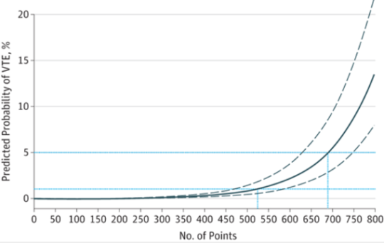
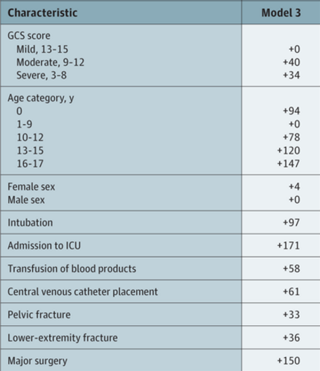
The tool consists of a list of risk factors, each with an assigned point value. The total point value is then identified on a chart of the regression equation, which shows the risk of VTE in percent.

Here are the factors:

Note that the highest risk factors are age >= 13, ICU admission, and major surgery.

And here is the regression chart:

Bottom line: This is a nice tool, and it’s time for some clinical validation. So now all we have to do is figure out how much risk is too much, and determine which prophylactic tools to use at what level. The key to making this clinically usable is to have a readily available “VTE Risk Calculator” available at your fingertips to do the grunt work. Hmm, maybe I’ll chat with the authors and help develop one!

Reference: A Clinical Tool for the Prediction of Venous Thromboembolism in Pediatric Trauma Patients. JAMA Surg 151(1):50-57, 2016.