Tag Archives: VTE

How Early Can We Start Chemoprophylaxis In TBI Patients?

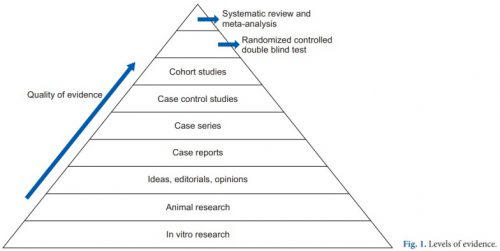
We’ve learned a couple of things in the last two posts by reviewing recent systematic review / meta-analysis studies. First, low molecular weight heparin provides better prophylaxis against venous thromboembolism (VTE) than unfractionated heparin. And giving prophylaxis within the first 72 hours of admission significantly decreases the incidence of VTE with no increase in existing intracranial bleeds or mortality.

So the only remaining question is, how low can you go? That is, how soon can you safely start chemoprophylaxis? The trauma group at George Washington University in DC put together a study to examine this question.

They, and one other Level I trauma center, performed a retrospective cohort study of adult, blunt TBI patients over a three year period. Patients with penetrating brain injury, and those with any other body region with significant injury (AIS >1) were excluded so this group truly represented isolated brain injury. Other exclusion criteria were progression of blood on CT within 6 hours, and crani or death within 24 hours. Early VTE prophylaxis was defined as occurring within 24 hours, and late was > 24 hours.

All patients had hourly neuro evaluations and a repeat head CT at six hours after admission. All had compression devices applied to their legs, and received either low molecular weight (LMWH) or unfractionated heparin (UH) at a fixed dose regarding of body habitus. Anti-Factor Xa levels were not measured.

Here are the factoids:

  • Between the two centers, 264 met inclusion criteria
  • About 40% received early prophylaxis and the remaining ones received their drug after 24 hours
  • ISS was higher (18 vs 15) and GCS was lower (13 vs 14) in the late therapy group
  • About 88% of patients in the early prophylaxis group received LMWH vs only 63% in the late group
  • Average time to prophylaxis start in the early group was 17 hours vs 47 hours in the late group
  • There were no differences in bleed progression between early and late groups (5.6% vs 7%)
  • The craniotomy / craniectomy rates were the same in early and late groups (1.9% vs 2.5%)
  • VTE rate was the same in early vs late groups (0% vs 2.5%)

Bottom line: The authors concluded that there was no additional risk in giving early VTE prophylaxis in TBI patients with a stable CT seven hours after arrival. This was true for patients with subdural, epidural, subarachnoid, and intraparenchymal bleeds.

But there are some limitations to consider. This was a retrospective study, and was a “how we do it” study” as well in terms of the choice of LMWH vs UH. This means there was not protocol for the form of heparin used; that was determined by surgeon preference. 

There was also a difference in ISS and GCS between groups. However, the difference may not have been clinically significant, and it could have made the late group look worse if it were. Statistically, it did not.

And finally, the numbers are small and there was no power analysis. So there is the question of whether a significant difference could have even been detected.

What does it all mean? Well, it suggests that early (within 24 hours) chemoprophylaxis does not cause harm compared to later administration. But the study is not definitive enough to change practice yet. It should definitely prompt discussions and practice guideline development for starting prophylaxis after 24 hours of CT scan stability now. And hopefully these authors (or others) are planning a better prospective study to help us start even sooner!

Reference: Early chemoprophylaxis against venous thromboembolism in patients with traumatic brain injury. Am Surgeon 88(2):187-193, 2021.

Unfractionated vs Low Molecular Weight Heparin For Trauma Patients

In my last post, I described some of the telltale signs that could be seen in a trauma center’s TQIP report that might suggest there are issues with how they go about providing prophylaxis for venous thromboembolism for their patients. Today, I will analyze a systematic review and meta-analysis of a collection of research that compared the efficacy and safety of unfractionated heparin (UFH) to low molecular weight heparin (LMWH) specifically for trauma patients.

First, it’s important to understand the concept of research quality. There is a huge amount of research published these days, and it varies considerably in how well it is designed, executed, and analyzed. Here is a diagram that illustrates the levels of quality and the volume of research published at each level. By quality, I mean the applicability to clinical treatment of actual humans. For this reason, test tube and animal research are low on the pyramid.

The research that most people consider to be the “gold standard” (randomized, controlled, double blind) is very close to the top. There is one class that, if conducted properly, may even be better. That is the systematic review and meta-analysis.

Most people have heard of meta-analysis, and it can be very good by itself. This combines lots of smaller studies into one larger one. However, it may hampered by the quality of the studies included in the meta-analysis. The tenet of “garbage in equals garbage out” certainly holds. But a systematic review takes that one step further.

The systematic review compiles all possible studies related to a small set of research questions, and usually concentrates on the ones with the highest quality research design. The quality of each of the studies is evaluated, and a meta-analysis is then performed on the best. Results are usually represented in a forest plot. This is an easy way to illustrate the estimated results from a number of studies that address the same question. There is also an entry that shows the relative strength of all of the studies combined. Here’s an example:

There are seven studies included, and each is displayed with its risk ratio (RR) and confidence interval (CI). The final diamond is the combined RR and CI for the entire group of studies. In the example above, note that most of the studies have CI bars that extend over the risk ratio = 1 line, meaning they may not be significant. But when taken together, the final risk ratio of the group is well under 1.0 and does not cross over it, denoting significance.

Let’s now apply this concept to a group of studies comparing UFH and LMWH for prevention of VTE for trauma patients. Based on keyword search, the authors identified 1,227 records for screening. Of those, only 40 were tentativley found to directly address the question. After in-depth analysis, only 12 were eligible for final review. For various reasons, only about 1 in 100 papers could be used to try to analyze the question. This always shocks me.

Here are the efficacy results. All are statistically significant, and all but mortality were stated with moderate certainty. The mortality number had low certainty due to the fact there were only three studies and confidence intervals were very wide.

  • Deep venous thrombosis: LMWH reduced by about 35% compared to UH
  • Pulmonary embolism: LMWH reduced by 44% although certainty was low
  • Any VTE: LMWH reduced by about 30%
  • Mortality: LMWH reduced by 56% (low to very low certainty)

Safety was also analyzed, including bleeding events, unexpected return to OR, heparin induced thrombocytopenia (HIT), and “any adverse events.” All of the Total Confidence Interval diamonds were situated on the risk ratio = 1 line, denoting no significant change when comparing LMWH vs UH.  However, quality of this data was noted to be low due to the quality of the individual studies. This means that we do not really know the answer to the safety question with any certainty yet.

Bottom line: This is one of the best summaries of our research on UH vs LMHW to date. It broadly reviewed the available literature and found only a small subset to analyze. It is clear that LMWH is superior for prevention of DVT and VTE overall. However, the impact on pulmonary embolism and death is still unclear.

As far as safety, the studies are still of quality that is too low to use for a decent analysis. Although this study did not detect any increase in complications, we still can’t say with any degree of certainty.

So what does it all mean? We have been using LMWH for decades now. Most likely, if there were regular complications like bleeding, unexpected return to OR, or HIT we would have definitely noticed it by now. Fortunately, we only have a few anecdotes and case reports to scare us off.

Overall, there is good support for the use of LMWH exclusively in most trauma patients. However, the prescribing provider should always assess patient factors that may suggest that UH might be better is a specific case. But remember that using UH trades an unclear/unlikely safety advantage for a recognized decrease in efficacy.

Reference: Efficacy and safety of low molecular weight heparin versus unfractionated heparin for prevention of venous thromboembolism in trauma patients. Ann Surgery 275(1):19-28, 2022.

VTE Prophylaxis And TBI

There has been a tremendous amount of gnashing of teeth regarding venous thromboembolism (VTE) prophylaxis in patients with blood in their head. This means any kind of blood: subarachnoid / epidural / subdural hematomas as well as intraparenchymal hemorrhage.

Trauma professionals have traditionally been hesitant to give any type of anticoagulant to a patient who has just bled, or who may be at risk for bleeding in the very near future. This becomes even more important in areas like the brain where management is a bit more difficult and adverse events can be devastating.

For this reason, our neurosurgical colleagues frequently like to steer the ship and dictate what type of VTE prophylaxis can be given, and when. Unfortunately, much of their advice may be driven by dogma and what they learned about the subject during their training. Having studied hundreds of TQIP reports over the past few years, I’ve learned to pick out hospitals that are relying on the advice of non-trauma surgeons to direct the prophylactic regimen.

Here are two dead giveaways that something is amiss. First, look at your TQIP report table titled “Pharmacologic VTE Prophylaxis Type.”

Compare the use of unfractionated heparin vs low molecular weight heparin (LMWH). This hospital has a huge variance from the norm compared to other comparable trauma centers. This means that “someone” is dictating its use for some subset of patients.

In my experience, this is typically a neurotrauma thing. Now take a look at the TQIP table titled “Pharmacologic VTE Prophylaxis.” Specifically, look at the “Severe TBI” cohort for time to VTE prophylaxis.

It is very clear that there is a significant delay to administering VTE prophylaxis to TBI patients. These two data points indicate that there is some reluctance to giving appropriate treatment to these patients.

The literature is clear that VTE prophylaxis is important in many trauma patients, including those with serious head injury. There are three questions that need to be answered to settle on optimal care:

  1. Which chemoprophylaxis is best, unfractionated or low molecular weight heparin?
  2. Is it better to give the selected agent earlier or later?
  3. If earlier is better, how early can we give it?

I will address each of these questions in this series of posts, focusing on neurotrauma patients. In order to try to toss out dogma, the literature I cite will be recent, no more than about two years old. So join me for battle next week as we have unfractionated vs low molecular weight heparin face off.

Thanks to Jim Sargent from Beth Israel Deaconess Medical Center for suggesting this topic.

Best Of EAST #6: How Long Does Risk For VTE Last After Spine Fracture?

Most trauma centers use an existing venous thromboembolism (VTE) guideline or have developed their own injury-specific one. These include risk factors, contraindications, specific agent, and dosing recommendations. But one thing most do not include is duration of prophylaxis!

The length of time a patient is at risk for VTE is not well delineated yet. The group at the University of Arizona decided to tackle this program using the National Readmission Database. This dataset is a comprehensive resource for critically analyzing patients who are discharged and readmitted, even for multiple occurrences. It covers 30 states and almost two thirds of the population.

The authors focused on VTE occurring during the first six months after injury. Patients who died on the initial admission, were taking anticoagulants, had spinal surgery, or sustained a spinal cord injury were excluded. Over 41,000 records from the year 2017 met these criteria.

Here are the factoids:

  • The average age was 61, which shows the skew toward the elderly with these injuries
  • Spine areas injured were cervical in 20%, thoracic in 19%, lumbar in 29%, sacrococcygeal in 11%, and multiple levels in 21%.
  • During the initial admission, 1.5% developed VTE: 0.9% were DVT and 0.7% were PE
  • Within 1 month of discharge, 0.6% of patients were readmitted for VTE: 0.4% DVT and 0.3% PE
  • In the first 6 months, 1.2% had been readmitted: 0.9% DVT and 0.6% PE
  • Mortality in the first 6 months was 6.7%
  • Factors associated with readmission for VTE included older age, discharge to a skilled nursing facility, rehab center, or care facility

The authors concluded that VTE risk remains high up to 6 months after conservatively managed spinal fractures. They recommend further study to determine the ideal prophylactic agent and duration.

Bottom line: This is a creative way of examining a difficult problem. We know that VTE risk does not stop when our patient is discharged. This is one of the few ways to get a sense of readmissions, even if it is not to the same hospital. And remember, this is an underestimate because it’s possible for a patient living near a state border to be re-hospitalized in a state not in this database.

This study might prompt us to prescribe up to six months of prophylaxis, particularly in seniors who are discharged to other care facilities.

Here are my questions for the author and presenter:

  • Is there any way to extrapolate your data to the entire population of the US, or to compensate for the “readmission over state lines” problem?
  • Is the odds ratio of 1.01 for risk of VTE in the elderly age group significant in any way? It seems like a very low number that would be easily overwhelmed by the “noise” in this data set.
  • Is the mortality number for all causes, or just VTE?

This is an intriguing study, and one that should influence the VTE guidelines in place at many trauma centers!

Reference: THE LONG-TERM RISKS OF VENOUS THROMBOEMBOLISM AFTER NON-OPERATIVELY MANAGED SPINAL FRACTURE. EAST 35th ASA, oral abstract #28.

Routine Duplex Screening For Venous Thromboembolism

Venous thromboembolism (VTE) is a potential problem for all hospitalized patients, and traumatic injury is yet an additional risk factor for its occurrence. Most trauma centers have some kind of risk assessment tool to help the tailor their chemoprophylaxis regimen to patients most at risk. But far fewer have adopted the use of screening ultrasounds to monitor for new onset VTE that would dictate conversion to therapeutic treatment.

Unfortunately, in the US, the Centers for Medicare and Medicaid Services (CMS) has deemed VTE as a “never” event and penalizes hospitals when they report it. One of the unintentional consequences of this (or is it?) is that hospitals may then pressure trauma programs to avoid surveillance in order to “make the numbers look better.” Remember Law X from Samuel Shem’s House of God?

X. If you don’t take a temperature, you can’t find a fever.

Similarly, if you don’t do a duplex screen, you probably won’t detect VTE. Now granted. some patients develop classic symptoms like edema, pain, and tenderness. But not that many.

But is this wise? My contention has been that if the patient doesn’t develop symptoms that catch your attention, yet they develop VTE that you don’t know about, they are at risk for more serious complications like pulmonary embolism (PE). And you are blithely unaware.

The trauma group at Intermountain Medical Center in Salt Lake City performed an elegant study to determine the impact of screening for VTE in their trauma patients. They performed a prospective, randomized trial on trauma patients admitted over a 30-month period. Patients were included if they were judged to be at moderate to high risk based on their risk assessment profile (RAP) score. Patients were excluded if they were children, had VTE or PE within 6 months prior to hospitalization, or had some type of hypercoagulable state.

Patients were sequentially randomized to no duplex screening vs screening on days 1, 3, 7, and then weekly thereafter. The primary outcome measure was PE during the hospital stay. Secondary outcomes consisted of a number of factors relating to development of DVT.

Here are the factoids:

  • Nearly two thousand patients were enrolled, with about 995 patients in each group and no differences in demographics
  • The ultrasound group had significantly more below-knee (124 vs 8) and above knee (19 vs 8) DVT identified (no surprise there)
  • The ultrasound group had significantly fewer pulmonary emboli than the no ultrasound group (1 vs 9) (lots of surprise here!)
  • Mortality was similar during the hospital stay and for 90 days after

Bottom line: If you look for it, you will find it! This is the definition of surveillance bias. But in in this study, looking for clots in the legs may also decrease the number of patients who develop symptomatic pulmonary embolism. How could this be?

There are a few possibilities. The majority of DVT found in the surveillance group were located distally. Although there is some uncertainty as to how likely these are to embolize, it is probably very low. So let’s ignore them for now and assume that only the proximal clots might embolize.

This leaves an extra 11 DVT found in the surveillance group over and above the no-ultrasound group. Despite that, the surveillance group had only one PE vs 9 in the no-ultrasound group!

Another explanation was that the ultrasound guided changes in management, shifting to management to therapeutic drug dosing. The authors did not find a significant difference between the use of therapeutic vs prophylactic dosing between the groups. But there was a difference. Although the overall study was well-powered, there really weren’t enough numbers to show whether there was a true difference in therapeutic dosing. Fourteen patients in the ultrasound group got therapeutic anticoagulation compared to only 4 in the no-surveillance group. I think this is the actual reason.

Overall, this is a well-designed and well-executed study that shows why taking the Ron Popeil approach to DVT prophylaxis (“set it and forget it”) doesn’t work. Patients do occasionally develop proximal DVT on standard chemoprophylaxis (and frequently develop distal DVT), but it doesn’t always result in obvious signs and symptoms. This study shows that if you don’t look for it, you may not know until they suddenly develop chest pain, air hunger, and worse! So consider carefully if your practice guideline doesn’t yet include surveillance.

Reference: Trauma Patients at Risk for Venous Thromboembolism who Undergo Routine Duplex Ultrasound Screening Experience Fewer Pulmonary Emboli: A Prospective Randomized Trial. J Trauma, publish ahead of print, Publish Ahead of Print. DOI: 10.1097/TA.0000000000003104, February 4, 2021.