Tag Archives: pulmonary embolism

Predicting VTE Risk In Children

There’s a lot of debate about if and at what age injured children develop significant risk for venous thromboembolism (VTE). In the adult world, it’s a little more clear cut, and nearly every patient gets some type of prophylactic device or drug. Kids, we’re not so certain about at all.

The Children’s Hospital of Wisconsin tried to tease out these factors to develop and implement a practice guideline for pediatric VTE prophylaxis. They prospectively reviewed over 4000 pediatric patients admitted over a 6 year period.

It looks like the guideline was developed using some or all of this data, then tested using regression models to determine which factors were significant. The guideline was then tweaked and a final model was implemented.

Here are the factoids:

  • 588 of the patients (14%) were admitted to the ICU, and 199 of these were identified as high-risk by the guidelines
  • Median age was 10 (this is always important in these studies)
  • VTE occurred in 4% of the ICU patients, and 10% of the high-risk ones
  • Significant risk factors included presence of central venous catheter, use of inotropes, immobilization, and GCS < 9

Bottom line: This abstract confuses me. How were the guidelines developed? What were they, exactly? And the results seem to pertain to the ICU patients only. What about the non-ICU kids? The abstract just can’t convey enough information to do the study justice. Hopefully, the oral presentation will explain all.

I prefer a very nice analysis done at the Oregon Health Science University in Portland. I wrote about this study earlier this year. The authors developed a very useful calculator that includes most of the risk factors in this model, and a few more. Input the specific risks, and out comes a nice score. The only issue is, what is the score threshold to begin prophylaxis and monitoring? Much more practical (and understandable) than this abstract. Check it out at the link below.

References:

  1. Evaluation of guidelines for injured children at high risk for venous thromboembolism: A prospective observational study. J Trauma Acute Care Surg. 2017 May;82(5):836-844.
  2. A Clinical Tool for the Prediction of Venous Thromboembolism in Pediatric Trauma Patients. JAMA Surg 151(1):50-57, 2016.

Unfractionated vs Low Molecular Weight Heparin For Trauma Patients

In my last post, I described some of the telltale signs that could be seen in a trauma center’s TQIP report that might suggest there are issues with how they go about providing prophylaxis for venous thromboembolism for their patients. Today, I will analyze a systematic review and meta-analysis of a collection of research that compared the efficacy and safety of unfractionated heparin (UFH) to low molecular weight heparin (LMWH) specifically for trauma patients.

First, it’s important to understand the concept of research quality. There is a huge amount of research published these days, and it varies considerably in how well it is designed, executed, and analyzed. Here is a diagram that illustrates the levels of quality and the volume of research published at each level. By quality, I mean the applicability to clinical treatment of actual humans. For this reason, test tube and animal research are low on the pyramid.

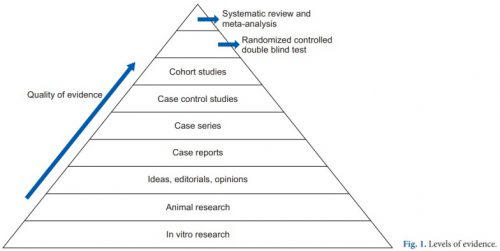
The research that most people consider to be the “gold standard” (randomized, controlled, double blind) is very close to the top. There is one class that, if conducted properly, may even be better. That is the systematic review and meta-analysis.

Most people have heard of meta-analysis, and it can be very good by itself. This combines lots of smaller studies into one larger one. However, it may hampered by the quality of the studies included in the meta-analysis. The tenet of “garbage in equals garbage out” certainly holds. But a systematic review takes that one step further.

The systematic review compiles all possible studies related to a small set of research questions, and usually concentrates on the ones with the highest quality research design. The quality of each of the studies is evaluated, and a meta-analysis is then performed on the best. Results are usually represented in a forest plot. This is an easy way to illustrate the estimated results from a number of studies that address the same question. There is also an entry that shows the relative strength of all of the studies combined. Here’s an example:

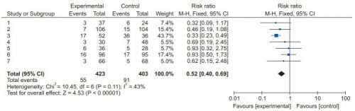
There are seven studies included, and each is displayed with its risk ratio (RR) and confidence interval (CI). The final diamond is the combined RR and CI for the entire group of studies. In the example above, note that most of the studies have CI bars that extend over the risk ratio = 1 line, meaning they may not be significant. But when taken together, the final risk ratio of the group is well under 1.0 and does not cross over it, denoting significance.

Let’s now apply this concept to a group of studies comparing UFH and LMWH for prevention of VTE for trauma patients. Based on keyword search, the authors identified 1,227 records for screening. Of those, only 40 were tentativley found to directly address the question. After in-depth analysis, only 12 were eligible for final review. For various reasons, only about 1 in 100 papers could be used to try to analyze the question. This always shocks me.

Here are the efficacy results. All are statistically significant, and all but mortality were stated with moderate certainty. The mortality number had low certainty due to the fact there were only three studies and confidence intervals were very wide.

  • Deep venous thrombosis: LMWH reduced by about 35% compared to UH
  • Pulmonary embolism: LMWH reduced by 44% although certainty was low
  • Any VTE: LMWH reduced by about 30%
  • Mortality: LMWH reduced by 56% (low to very low certainty)

Safety was also analyzed, including bleeding events, unexpected return to OR, heparin induced thrombocytopenia (HIT), and “any adverse events.” All of the Total Confidence Interval diamonds were situated on the risk ratio = 1 line, denoting no significant change when comparing LMWH vs UH.  However, quality of this data was noted to be low due to the quality of the individual studies. This means that we do not really know the answer to the safety question with any certainty yet.

Bottom line: This is one of the best summaries of our research on UH vs LMHW to date. It broadly reviewed the available literature and found only a small subset to analyze. It is clear that LMWH is superior for prevention of DVT and VTE overall. However, the impact on pulmonary embolism and death is still unclear.

As far as safety, the studies are still of quality that is too low to use for a decent analysis. Although this study did not detect any increase in complications, we still can’t say with any degree of certainty.

So what does it all mean? We have been using LMWH for decades now. Most likely, if there were regular complications like bleeding, unexpected return to OR, or HIT we would have definitely noticed it by now. Fortunately, we only have a few anecdotes and case reports to scare us off.

Overall, there is good support for the use of LMWH exclusively in most trauma patients. However, the prescribing provider should always assess patient factors that may suggest that UH might be better is a specific case. But remember that using UH trades an unclear/unlikely safety advantage for a recognized decrease in efficacy.

Reference: Efficacy and safety of low molecular weight heparin versus unfractionated heparin for prevention of venous thromboembolism in trauma patients. Ann Surgery 275(1):19-28, 2022.

Routine Duplex Screening For Venous Thromboembolism

Venous thromboembolism (VTE) is a potential problem for all hospitalized patients, and traumatic injury is yet an additional risk factor for its occurrence. Most trauma centers have some kind of risk assessment tool to help the tailor their chemoprophylaxis regimen to patients most at risk. But far fewer have adopted the use of screening ultrasounds to monitor for new onset VTE that would dictate conversion to therapeutic treatment.

Unfortunately, in the US, the Centers for Medicare and Medicaid Services (CMS) has deemed VTE as a “never” event and penalizes hospitals when they report it. One of the unintentional consequences of this (or is it?) is that hospitals may then pressure trauma programs to avoid surveillance in order to “make the numbers look better.” Remember Law X from Samuel Shem’s House of God?

X. If you don’t take a temperature, you can’t find a fever.

Similarly, if you don’t do a duplex screen, you probably won’t detect VTE. Now granted. some patients develop classic symptoms like edema, pain, and tenderness. But not that many.

But is this wise? My contention has been that if the patient doesn’t develop symptoms that catch your attention, yet they develop VTE that you don’t know about, they are at risk for more serious complications like pulmonary embolism (PE). And you are blithely unaware.

The trauma group at Intermountain Medical Center in Salt Lake City performed an elegant study to determine the impact of screening for VTE in their trauma patients. They performed a prospective, randomized trial on trauma patients admitted over a 30-month period. Patients were included if they were judged to be at moderate to high risk based on their risk assessment profile (RAP) score. Patients were excluded if they were children, had VTE or PE within 6 months prior to hospitalization, or had some type of hypercoagulable state.

Patients were sequentially randomized to no duplex screening vs screening on days 1, 3, 7, and then weekly thereafter. The primary outcome measure was PE during the hospital stay. Secondary outcomes consisted of a number of factors relating to development of DVT.

Here are the factoids:

  • Nearly two thousand patients were enrolled, with about 995 patients in each group and no differences in demographics
  • The ultrasound group had significantly more below-knee (124 vs 8) and above knee (19 vs 8) DVT identified (no surprise there)
  • The ultrasound group had significantly fewer pulmonary emboli than the no ultrasound group (1 vs 9) (lots of surprise here!)
  • Mortality was similar during the hospital stay and for 90 days after

Bottom line: If you look for it, you will find it! This is the definition of surveillance bias. But in in this study, looking for clots in the legs may also decrease the number of patients who develop symptomatic pulmonary embolism. How could this be?

There are a few possibilities. The majority of DVT found in the surveillance group were located distally. Although there is some uncertainty as to how likely these are to embolize, it is probably very low. So let’s ignore them for now and assume that only the proximal clots might embolize.

This leaves an extra 11 DVT found in the surveillance group over and above the no-ultrasound group. Despite that, the surveillance group had only one PE vs 9 in the no-ultrasound group!

Another explanation was that the ultrasound guided changes in management, shifting to management to therapeutic drug dosing. The authors did not find a significant difference between the use of therapeutic vs prophylactic dosing between the groups. But there was a difference. Although the overall study was well-powered, there really weren’t enough numbers to show whether there was a true difference in therapeutic dosing. Fourteen patients in the ultrasound group got therapeutic anticoagulation compared to only 4 in the no-surveillance group. I think this is the actual reason.

Overall, this is a well-designed and well-executed study that shows why taking the Ron Popeil approach to DVT prophylaxis (“set it and forget it”) doesn’t work. Patients do occasionally develop proximal DVT on standard chemoprophylaxis (and frequently develop distal DVT), but it doesn’t always result in obvious signs and symptoms. This study shows that if you don’t look for it, you may not know until they suddenly develop chest pain, air hunger, and worse! So consider carefully if your practice guideline doesn’t yet include surveillance.

Reference: Trauma Patients at Risk for Venous Thromboembolism who Undergo Routine Duplex Ultrasound Screening Experience Fewer Pulmonary Emboli: A Prospective Randomized Trial. J Trauma, publish ahead of print, Publish Ahead of Print. DOI: 10.1097/TA.0000000000003104, February 4, 2021.

Best Of EAST #8: Early vs Late Full Anticoagulation In TBI

Trauma professionals are always reluctant to anticoagulate TBI patients with demonstrated blood in their head. In recent years, we’ve become more comfortable providing prophylactic doses of low molecular weight heparin after a suitable period. This is typically 24-48 hours after a stable head CT in patients with select types of intracranial hemorrhage (ICH) who are at increased risk for venous thromboembolism.

But what about therapeutic dose anticoagulation in these patients? Let’s say that you have a patient with ICH who has developed a significant pulmonary embolism (PE)? Is is safe to give full dose anticoagulation? And if so, when?

The group at Shock Trauma in Baltimore attempted to answer this in one of the EAST Quick Shot presentations scheduled for this week. The did a retrospective review of 4.5 years of their own data on these patients. They specifically selected patients who had both ICH and PE and compared those who received full anticoagulation within 7 days of injury vs those who were dosed after 7 days. Outcomes studied included death, interventions for worsening ICH, and pulmonary complications.

Here are the factoids:

  • A total of 50 patients had both ICH and PE, but only the 46 who received therapeutic anticoagulation were analyzed
  • 19 patients (41%) received early anticoagulation, and 27 received it late (59%)
  • There were 4 deaths in the early group (2 from the PE, 1 from multi-system organ failure, 1 from the TBI) vs none in the late group, and this was statistically significant
  • 3 patients in the early group (18%) vs 2 in the late group (7%) had an increase in their ICH (p=0.3), and none required intervention

The authors concluded that their study failed to show any instances of clinically significant progression of ICH after anticoagulation, and that it is not associated with worse outcomes, even if started early. Thus they recommend that ICH should not preclude full anticoagulation, even early after injury.

My comment: I always say that you shouldn’t let one paper change your practice. Even a really good one. In order to ensure that you are providing the best care, more work must always be done to confirm (or refute) the findings of any provocative research. And this little Quick Shot, with little opportunity for questions from the audience, should definitely not change it!

The major issues to consider here are common ones: 

  • This was a retrospective study and it does not appear that any guideline was followed to determine who got early vs late anticoagulation. So who knows what kind of selection bias was occurring and how the surgeon decided to prescribe anticoagulation? It’s very possible that patients with a “bad CT” were put into the late group, and the not so bad ones in the early group. This would bias the results toward better outcomes in the early anticoagulation group.
  • It’s also a very small study that is extremely underpowered. The authors comment on the fact that the outcomes of the early group were not worse than the late group. However, looking at their sample size (46) shows that they would only be able to show differences if they were about 5x worse in the early group. They would realistically need about 350 total patients to truly show that the groups behaved the same. Their small numbers also preclude saying that there were no ICH progressions. There very well could have been if 300 more patients were added to the series.
  • And isn’t death a significant outcome? The authors indicated that 2 of the 4 deaths were a result of the PE. Yet there was a significant association (p=0.02) of increased death in the early anticoagulation patients that can’t be discounted.

Bottom line: It’s way too early to consider giving early anticoagulation to patients with ICH and pulmonary embolism. It may very well be shown to be acceptable, eventually. But not yet. And a much bigger prospective study will be required to confirm it.

Reference: Therapeutic anticoagulation in patients with traumatic brain injuries and pulmonary emboli. EAST Annual Assembly Quick Shot #7, 2020.

The IVC Filter In Trauma: Why?

The inferior vena cava (IVC) filter has been around in one form or another for over 40 years. One would think that we would have figured everything about it out by now. But no!  The filter has evolved through a number of iterations and form factors over the years. The existing studies, in general, give us piecemeal information on the utility and safety of the device.

One of the major innovations with this technology came with the development of a removable filter. Take a look at the product below. Note the hook at the top and the (relatively) blunt tips of the feet. This allows a metal sheath to be slipped over the filter while in place in the IVC. The legs collapse, and the entire thing can be removed via the internal jugular vein.

ivc-filter-complications1

The availability of the removable filter led the American College of Chest Physicians to recommend their placement in patients with known pulmonary embolism (PE) or proximal deep venous thrombosis (DVT) in patients with contraindications to anticoagulation. Unfortunately, this has been generalized by some trauma professionals over the years to include any trauma patients at high risk for DVT or PE, but who don’t actually have them yet.

One would think that, given the appearance of one of these filters, they would be protective and clots would get caught up in the legs and be unable to travel to the lungs as a PE. Previous studies have taught us that this is not necessarily the case. Plus, the filter can’t stop clots that originate in the upper extremities from becoming an embolism. And there are quite a few papers that have demonstrated the short- and long-term complications, including clot at and below the filter as well as post-phlebitic syndrome in the lower extremities.

A study from Boston University reviewed their own experience retrospectively over a 9 year period. This cohort study looked at patients with and without filters, matching them for age, sex, race, and injury severity. The authors specifically looked at mortality, and used four study periods during the 9 year interval.

Here are the factoids:

  • Over 18,000 patients were admitted during the study period, resulting in 451 with an IVC filter inserted and 1343 matched controls
  • The patients were followed for an average of 4 years after hospitalization
  • Mortality was identical between patients with filters vs the matched controls

dvt-study

  • There was still no difference in mortality, even if the patients with the filter had DVT or PE present when it was inserted
  • Only 8% ever had their “removable” filter removed (!)

Bottom line: Hopefully, it’s becoming obvious to all that the era of the IVC filter has come and gone. There are many studies that show the downside of placement. And there are several (including this one) that show how forgetful we are about taking them out when no longer needed. And, of course, they are expensive. But the final straw is that they do not seem to protect our patients like we thought (hoped?) they would. It’s time to reconsider those DVT/PE protocols and think really hard about whether we should be inserting IVC filters in trauma patients at all.

Tomorrow: a look at trends in filter insertion and retrieval.

Related post:

Reference: Association Between Inferior Vena Cava Filter Insertion
in Trauma Patients and In-Hospital and Overall Mortality. JAMA Surg, online ahead of print, September 28, 2016.