Category Archives: CNS

Best Of AAST 2022 #5: Traumatic Subarachnoid Hemorrhage

Traditionally, just about any type of intracranial hemorrhage (ICH) prompts a thorough neurological investigation and frequently involves a neurosurgery consultation. Subarachnoid hemorrhage (SAH) is one type of ICH that has historically demonstrated very few adverse consequences. Yet the custom of performing a time-consuming and expensive evaluation has persisted.

Over the past few years, the literature about the lack of utility of this practice has been growing. Many trauma centers (including my own at Regions Hospital) have simplified the evaluation of traumatic SAH. We have actually come to a point that we don’t even admit most of these patients. And if we are not going to admit them, why should we even accept them in transfer from other hospitals?

The trauma group at Lehigh Valley in Pennsylvania performed a five-year review of their own hospital experience (Level I trauma center) from 2015-2019. They wanted to answer the question about the necessity of transferring SAH patients to a higher-level trauma center for evaluation. They limited the study to patients with an isolated SAH. These patients were evaluated for factors that would indicate the potential for a worsening repeat CT scan.

Here are the factoids:

  • The study patients were older (mean 73 years) with a slight preponderance of females
  • ISS was low (mean 5) as would be expected for an isolated injury
  • Slightly more than half (57%) were transferred from other hospitals
  • A total of 350 patients were reviewed, and 97% had a neurosurgery consult
  • Only one patient required neurosurgical intervention, and four died in hospital
  • A total of 311 patients had a repeat CT scan and 16 showed a worse result
  • Higher AIS head was associated with a worse result

The authors concluded that it is safe to manage traumatic SAH without transfer to a higher level trauma center.  They also suggest that neurosurgical consultation is not needed and that repeat imaging may not be very useful.

Bottom line: This abstract conforms to my own opinion, so I have to take great care not to succumb to my confirmation bias. The authors found that nearly no one needed neurosurgical intervention and concluded that a neurosurgical consult is probably not needed. This is all based on a low incidence of progression and few adverse events. I agree totally.

But I also think that referring hospitals will find these few adverse events and progressions troubling. Remember, 16 patients had a worse scan,  four died during their hospital stay, and one needed an operation. Thus, it will be very important for the authors to explain the details of these patients to assuage any fears that they might deteriorate in the outside hospital ED. 

My own experience indicates that “worsening CT” is typically just a slightly larger SAH and not the sudden development of subdural or epidural blood. This is more likely to happen in patients on thinners and they should be excluded from this pool anyway. They have very specific evaluation needs.

Based on recent published literature and our own clinical experience, the Regions trauma program discharges any patient with traumatic SAH who is not on thinners and has a GCS 15. They are given instructions to follow up with their primary care provider, and to schedule a TBI clinic appointment if they feel they have any persistent post-concussive symptoms. And because of this practice, the ED will not accept transfer of patients with this isolated injury.

You can download a copy of the protocol here.

Here are my questions for the authors and presenter:

  1. Please provide details on the patients with CT progression, operation, and death. We need to know if these were serious CT changes, or just incidental findings and complications from other causes.
  2. How successful have you been at resisting transfers in? Most referring centers are initially “not comfortable” with these patients, even though they can easily follow our simple evaluation guideline. It takes time and education to bring them into the fold.

I enjoyed this abstract and await the details to come in the presentation!

Print Friendly, PDF & Email

Epidural Hematoma Treated With Middle Meningeal Artery Embolization

Epidural hematoma is a life-threatening condition that is typically associated with arterial bleeding outside of the dura. Most frequently, this is due to a skull fracture that extends across and lacerates the middle meningeal artery (MMA).

The standard treatment regimen is neurologic monitoring in patients who have a (nearly) normal GCS and do not change neurologically. That escalates to rapid craniectomy and evacuation in those with neurologic compromise.  Interestingly, there have been a few case reports over the last 10 years describing attempted management by embolization of the MMA.

Let’s look at this idea more critically. This seems like it should be a good idea. But remember, in medicine you’ve actually got to study it. There are too many examples of things that make sense that are worthless or actually cause harm.

The first report I found was a series of one in which the patient was found to have a large subdural hematoma. He was taken to surgery and the lesion was evacuated. However, there was persistent epidural bleeding intraop which was thought to be controlled. Repeat scan the next day showed a large epidural, so he was returned to the OR. Once again, there was persistent epidural oozing and the collection was removed. Followup CT showed yet another epidural. The patient was finally taken to interventional radiology for embolization of the MMA. This was successful, and the patient had no further recurrences.

This case provided proof of concept, although the bleeding was not due to known traumatic injury to the MMA. Last year, another case report was published that described an experience (of one again) with a young male who was found down. He awakened and then became obtunded again. CT showed bilateral epidural hematomas. He was taken to the OR for operative evacuation of the larger one. Postop CT showed expansion of the smaller one.

The patient was then taken to the endovascular suite and MMA embolization was carried out. The hematoma stabilized and the patient was later discharged without sequelae.

This case was trauma-related, but not for an acute bleed. Now, let’s look at a bigger case series to see how well this works. This one detailed the experience of a neurosurgery group in Sao Paulo, Brazil. All patients who underwent conservative management based on “standard criteria” were studied. Patients with large hematomas, midline shift, depressed skull fracture, coagulopathy, or incomplete data were excluded. One third of the injuries were due to falls, and the rest were due to other blunt mechanisms.

Here are the factoids:

  • 85% had an attendant skull fracture
  • About 82% had active extravasation from the MMA
  • All patients had followup CT scan 1-7 days after the procedure, and no increase in epidural size was noted
  • None of the patients had a change in GCS or needed operative intervention
  • The authors compared these results to historical controls from other published literature

Bottom line: Sounds impressive, right? But not so fast, there are a lot of loose ends here. First, these are supposedly all patients with epidural hematoma who were treated without operation. Decision to operate was based on criteria set out in a paper published 15 years ago. This strains the imagination a bit. There is usually no uniformity in the way individual neurosurgeons decide to operate, so it is likely there may be some significant selection bias here. It is very easy to believe that patients who were predicted to do well were the only ones enrolled in the study. This also explains why the authors had to use controls from other authors’ research for outcome comparison.

The results are too clean as well. No adverse events. No patients who ended up needing surgery. Followup scans were performed any time between postop day 1 and 7, but there is no frequency breakdown. If most of the repeat scans were performed near the beginning of the postop period, little change would be expected. MMA embolization is either a miracle cure or …

You know what they say, “if it seems to good to be true…” A single case series like this should never change one’s practice. Middle meningeal artery embolization sounds like common sense, but the devil is always in the details. This concept needs a lot more study before you should ever consider it in your patients. Or, you could start a real, IRB-approved study and make an excellent contribution to the neurosurgery literature.

References:

  1. Embolization of the Middle Meningeal Artery for the Treatment of Epidural Hematoma. J Neurosurg 110(6):1247-1249, 2009.
  2. Middle Meningeal Artery Embolization for the Treatment of an Expanding Epidural Hematoma. World Neurosurg 128:284-286,2019
  3. Endovascular Management of Acute Epidural Hematomas: Clinical Experience With 80 Cases. J Neurosurg 128(4):1044-1050, 2018.
Print Friendly, PDF & Email

Best Of EAST #8: Early vs Late Full Anticoagulation In TBI

Trauma professionals are always reluctant to anticoagulate TBI patients with demonstrated blood in their head. In recent years, we’ve become more comfortable providing prophylactic doses of low molecular weight heparin after a suitable period. This is typically 24-48 hours after a stable head CT in patients with select types of intracranial hemorrhage (ICH) who are at increased risk for venous thromboembolism.

But what about therapeutic dose anticoagulation in these patients? Let’s say that you have a patient with ICH who has developed a significant pulmonary embolism (PE)? Is is safe to give full dose anticoagulation? And if so, when?

The group at Shock Trauma in Baltimore attempted to answer this in one of the EAST Quick Shot presentations scheduled for this week. The did a retrospective review of 4.5 years of their own data on these patients. They specifically selected patients who had both ICH and PE and compared those who received full anticoagulation within 7 days of injury vs those who were dosed after 7 days. Outcomes studied included death, interventions for worsening ICH, and pulmonary complications.

Here are the factoids:

  • A total of 50 patients had both ICH and PE, but only the 46 who received therapeutic anticoagulation were analyzed
  • 19 patients (41%) received early anticoagulation, and 27 received it late (59%)
  • There were 4 deaths in the early group (2 from the PE, 1 from multi-system organ failure, 1 from the TBI) vs none in the late group, and this was statistically significant
  • 3 patients in the early group (18%) vs 2 in the late group (7%) had an increase in their ICH (p=0.3), and none required intervention

The authors concluded that their study failed to show any instances of clinically significant progression of ICH after anticoagulation, and that it is not associated with worse outcomes, even if started early. Thus they recommend that ICH should not preclude full anticoagulation, even early after injury.

My comment: I always say that you shouldn’t let one paper change your practice. Even a really good one. In order to ensure that you are providing the best care, more work must always be done to confirm (or refute) the findings of any provocative research. And this little Quick Shot, with little opportunity for questions from the audience, should definitely not change it!

The major issues to consider here are common ones: 

  • This was a retrospective study and it does not appear that any guideline was followed to determine who got early vs late anticoagulation. So who knows what kind of selection bias was occurring and how the surgeon decided to prescribe anticoagulation? It’s very possible that patients with a “bad CT” were put into the late group, and the not so bad ones in the early group. This would bias the results toward better outcomes in the early anticoagulation group.
  • It’s also a very small study that is extremely underpowered. The authors comment on the fact that the outcomes of the early group were not worse than the late group. However, looking at their sample size (46) shows that they would only be able to show differences if they were about 5x worse in the early group. They would realistically need about 350 total patients to truly show that the groups behaved the same. Their small numbers also preclude saying that there were no ICH progressions. There very well could have been if 300 more patients were added to the series.
  • And isn’t death a significant outcome? The authors indicated that 2 of the 4 deaths were a result of the PE. Yet there was a significant association (p=0.02) of increased death in the early anticoagulation patients that can’t be discounted.

Bottom line: It’s way too early to consider giving early anticoagulation to patients with ICH and pulmonary embolism. It may very well be shown to be acceptable, eventually. But not yet. And a much bigger prospective study will be required to confirm it.

Reference: Therapeutic anticoagulation in patients with traumatic brain injuries and pulmonary emboli. EAST Annual Assembly Quick Shot #7, 2020.

Print Friendly, PDF & Email

AAST 2019 #5: DOACs Part 2

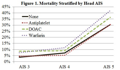
In my last post, I reviewed a study that scrutinized reversal of direct oral anticoagulants (DOACs), and the outcomes of using various reversal agents. Today I’ll look at an abstract that compared in-hospital outcomes of elderly patients with severe TBI who were taking a variety of anticoagulant drugs, including DOACs.

The group at St. Joseph Mercy Hospital in Ann Arbor reviewed the dataset from the Michigan Trauma Quality Improvement Program database over a seven year period. To be included, patients needed to be at least 65 years old, suffer a fall, and have a significant head injury (AIS > 3). The final data consisted of records from 8312 patients treated at both Level I and II trauma centers across the state.

Here are the factoids:

  • 40% of patients were taking antiplatelet agents, 13% warfarin, 4% DOAC, and the remaining half or so were taking nothing.
  • The head injuries were severe, with mean AIS of 4.
  • After adjusting for “patient factors”, mortality or hospital outcomes were 1.6x more likely when warfarin was used
  • Complication risk increased 1.4x for warfarin and 1.3x for antiplatelet patients, but not for DOACs
  • Hospital length of stay was a day longer in the warfarin group (6.7 days) vs about 5.7 in the others

The authors concluded that elderly patients with severe TBI on DOACs fared better than those on warfarin. They stated that this could help alleviate concerns about DOACs in head trauma patients.

This is yet another interesting and surprising piece of the TBI on anticoagulants puzzle! It is obviously limited due to its retrospective database nature, which prevents us from asking even more interesting questions of this dataset. And it completely prevents us from looking at the specifics of each case including decision making, imaging, etc. But it’s a good start that should prompt us to find even better sources of data to tease out the details we must know in order to improve this patient group’s care.

Here are my questions for the presenter and authors:

  • I am very interested in the “patient factors” that were adjusted for to try to normalize the groups. Please describe in detail the specific ones that were used so we can understand how this influenced your results.
  • This information is intriguing, suggesting that warfarin is more evil that DOACs. What is the next step? What shall we do to further elucidate the problems, and how can we ameliorate the mortality and complication effects?

This is more good stuff about DOACs, and I can’t wait to hear the details.

Print Friendly, PDF & Email

Redefining Mild TBI: Who Needs To Be Transferred?

One of the more common reasons for transfer to a higher level trauma center these days is the “mild or minimal TBI.” Technically, this consists of any patient with a Glasgow Coma Scale score of 14 or 15. A transfer is typically requested for observation or neurosurgical consultation, or because the clinicians at the initial hospital are not comfortable looking after the patient.

Is this really necessary? With the number of ground level falls approaching epidemic proportions, transferring all these patients could begin to overwhelm the resources of high level trauma centers. The surgical group at Carolinas Medical Center examined their experience with a simple scoring system they designed to predict high risk minor TBI patients, and thus suitability for transfer. Here is their checklist:

Category A
  • Traumatic SAH
  • Tentorial or falcine SDH < 4mm thickn
  • Convexity SDH < 4mm thick
  • Solitary IPH < 1cm
  • Isolated intraventricular hemorrhage < 4mm
Category B
  • Any Category A lesion greater than the allowed size
  • Midline shift
  • Skull fracture
  • Compression of basal cisterns
  • Diffuse SAH or SAH involving basal cisterns
  • Subacute or chronic SDH
SAH = subarachnoid hemorrhage, SDH = subdural hemorrhage, IPH = intraparenchymal hemorrhage

Patients were considered to be low risk if they had only one or two category A lesions. If they had more than two, or any Category B lesions, they were higher risk and transfer was considered justified.

The authors retrospectively reviewed their experience with these patients over a three year period. They followed patients to see if they needed neurosurgical intervention, and evaluated the cost savings of avoiding selective transfers based on their criteria.

Here are the factoids:

  • A total of 2120 patients were studied, with 68% low risk and 32% high risk
  • Two of the low risk patients (0.14%)  ultimately required neurosurgical intervention, compared to 21% of high risk patients
  • Mean age (56), and patients taking anticoagulants or antiplatelet agents were the same in the two groups, about 2-3% for each
  • System saving by avoiding EMS transfer costs would have been $734K had the low risk patients been kept at the initial hospital

Bottom line: This study was presented as a Quick Shot paper at this year’s Eastern Association for the Surgery of Trauma meeting, so there are some key details missing. Was there an association between anticoagulation or antiplatelet agent and two failures in the low risk group? What were they, and what intervention did they require?

If this data holds up to publication, then it may provide a useful tool for deciding to keep minimal TBI patients at the local hospital. This is usually far more convenient for the patient and their family, but would require additional education of the clinicians at that hospital to help them become comfortable managing these patients. 

We use a similar tool within our Level I trauma center to decide which patients require a neurosurgical consultation. Since the low risk patients almost never require intervention, our trauma service provides comprehensive management while in hospital, and arranges for TBI clinic followup post-discharge. You can view and download a copy using the link below.

Link: Regions Hospital SAH/IPH/Skull fracture practice guideline

Reference: Redefining minimal traumatic brain injury (MTBI): a novel CT criteria to predict intervention. Quick Shot Paper #48, EAST 2019.

Print Friendly, PDF & Email