Category Archives: CNS

GCS At 40: The Original GCS

The Glasgow Coma Score (GCS) has been in use for more than 40 years. Since that 40th anniversary a few years back, there has been talk of updating this tried and true system. But where did this scale come from? How was it devised? And why are we looking to update it now? I’ll dig into this topic over my next several posts.

The original paper describing the GCS was published in 1974 by Graham Teasdale and Bryan Jennett. They were neurosurgeons at the Institute of Neurologic Sciences in Glasgow, Scotland (of course) and were based in the Southern General Hospital. Until this paper was published, each report in the literature described its own assessment of level of consciousness. Most divided the spectrum into various steps noted between fully alert and comatose. Unfortunately, these systems were confusing, and they varied from 3-17 steps! There was just no consensus. Some relied on a comprehensive neurologic exam, including brainstem function tests. However, none of these were really designed for repeated bedside assessment.

Teasdale and Jennett settled on three simple areas to examine: eye-opening, motor response, and verbal response. They selected easily observable responses for each of these components. Here is a copy of the original scale:

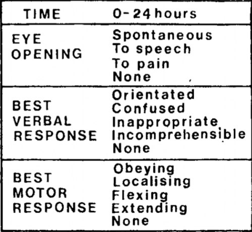
Notice that this differs from the current-day score. The motor response did not have a “withdrawal” option, so the maximum score was only 14! But that didn’t matter much at the time; the individual components were graphed out over time for inspection. A total score was not generally calculated.

Teasdale and Jennett found that inter-rater reliability for this system was excellent, compared to a 25% discrepancy for other less objective systems in use at the time. This led to its rapid adoption over the coming years.

In my next post, I’ll describe how GCS came to be used over the ensuing years.

Print Friendly, PDF & Email

Using MRI To Predict Outcome From Diffuse Axonal Injury (DAI)

Has this happened to you? A patient with a serious head injury is not waking up as expected. There were a few punctate hemorrhages seen on the initial CT scan. Your neurosurgery colleague orders an MRI to “provide a prognosis on the patient’s injury.”

Is this a legitimate request? Sure, MRI is very sensitive at detecting very small hemorrhages that may signal the presence of diffuse axonal injury (DAI). But do more abnormalities on MRI equal a poorer prognosis or longer recovery time?

A group from Vanderbilt presented their data from a retrospective cohort study at EAST earlier this year.  They reviewed 7 years of data from 2006 to 2012, including all patients with a head CT positive for intracranial injury and an MRI within 2 weeks. They excluded penetrating injuries and patients with psychiatric or neurologic disorders. They analyzed information on three year mortality, functional outcome, and quality of life.

Here are the factoids:

  • A total of 311 patients met all inclusion/exclusion criteria, with a median age of 40 and serious injury (average ISS 29, average ICU length of stay 6 days)
  • Functional status at discharge could be assessed in 240 patients, and only 118 could be contacted for long-term followup questions
  • Only 56% of patients with severe TBI had an MRI positive for DAI
  • Functional status was lower on discharge for patients positive for DAI on MRI
  • There was no difference in Glasgow Outcome Score, quality of life, or 3 year survival in patients with MRI evidence of DAI compared to those without

Bottom line: This is a relatively large study, but there are still several weaknesses that could skew the numbers a bit. However, it appears that MRI for prognostication of outcomes in patients with clinical DAI is not very helpful. First, only about half with a clinical picture of DAI showed it on MRI. And sure, MRI may tell us a little bit about their status when they are discharged from the hospital to rehab or transitional care. But is that information very useful? It certainly does not help predict their outcome in the longer term. So why order an expensive and difficult study (think restraints, sedation, lots of pumps and monitors) to tell us what we already know based on our experience with severe TBI?

Reference: Prognosis of diffuse axonal injury with traumatic brain injury. J Trauma 85(1):155-159, 2018

Print Friendly, PDF & Email

Sports Drinks And Electrolyte Replacement In TBI

Yesterday, I wrote about the (lack of) effectiveness of forcing hypernatremia in the management of TBI. However, we do know that some of our head injured patients have trouble maintaining a normal sodium level, and if it drops quickly or too far, hyponatremia can certainly cause problems. Trauma professionals have a number of tools to help fix this, including salt supplements or tablets, saline infusions, or even hypertonic saline in more difficult cases.

But what about using a sports drink to replace electrolytes? Isn’t that what athletes do? There are quite a few of these sports drinks on the market, and new ones seem to appear every week. Common examples are Gatorade, Powerade, Muscle Milk, Vitamin Water, 10-K Thirst Quencher, and many more. What if your brain injured patients eschews the salt tabs and insists on pounding down sports drinks all day?

Here is a table from an old sports medicine paper that describes the composition of a number of sports drinks from back in the day. Some, like Gatorade, are still around. (Click image to see a bigger, readable version)

Note that the electrolyte results are in mg/250cc, so I will translate to meq/liter for you. Gatorade had the highest sodium concentration at the time, 20meq/L, and one of the lowest potassiums at 3meq/L. The majority of the current day sports drinks have about the same electrolyte composition. Note that they are all a bit hyperosmolar (300+ mOsm), and this is made possible by added carbohydrate from some type of sugar. The carb is usually in the form of sucrose, dextrose, and/or high fructose corn syrup (yum!).

Bottom line: Your typical sports drink is equivalent to D30 in 0.1 normal saline. Not good for your TBI patient when consumed for sodium supplementation. It will actually drive the serum sodium down when consumed in quantity. Make sure your patients steer clear of this stuff until their brain injury is healed and they are running their next marathon.

Reference: The Effectiveness of Commercially Available Sports Drinks. Sports Med 29(3):181-209, 2000.

Print Friendly, PDF & Email

Targeted Hypernatremia In Trauma Brain Injury: Does This Work?

Traumatic brain injury (TBI) frightens and confuses most trauma professionals. The brain and its workings are a mystery, and there is very little real science behind a lot of what we do for TBI. One thing that we do know is that intracranial hypertension is bad. And another is that we do have some potent drugs (mannitol, hypertonic saline) to treat it emergently.

So if we can “dry out” the brain tissue on a moment’s notice and drop the ICP a bit with a hit of sodium, doesn’t it stand to reason that elevating the sodium level constantly might keep the brain from becoming edematous in the first place? Many neurosurgeons buy into this, and have developed protocols to maintain serum sodium levels in the mid-140s and higher. But what about the science?

A nice review was published in Neurocritical care which identified the 3 (!) papers that have promoted this practice in humans with TBI. In general, there was a decrease in ICP in the patients in the cited papers. Unfortunately, there were also a number of serious and sometimes fatal complications, including pulmonary edema and renal failure requiring hemodialysis. These complications generally correlated with the degree of hypernatremia induced. Papers were also reviewed that involved patients with other brain injury, not caused by trauma. Results were similar.

Bottom line: There is no good literature support, standard of care, or even consensus opinion for prophylactically inducing hypernatremia in patients with TBI. The little literature there is involves patients with severe TBI and ICP monitors in place. There is nothing written yet that justifies the expense (ICU level care) and patient discomfort (frequent blood draws) of using this therapy in patients with milder brain injury and a reliable physical exam. If you want to try out this relatively untried therapy, do us all a favor and design a nice study to show that the benefits truly outweigh the risks. 

And if you can point me to some supportive literature that I’ve missed, please do so!

Related posts:

References:

  • Induced and sustained hypernatremia for the prevention and treatment of cerebral edema following brain injury. Neurocrit Care 19:222-231, 2013.
  • Continuous hyperosmolar therapy for traumatic brain injury-induced cerebral edema: as good as it gets, or an iatrogenic secondary insult? J Clin Neurosci 20:30-31, 2013.
  • Continuous hypertonic saline therapy and the occurrence of complications in neurocritically ill patients. Crit Care Med 37(4):1433-1441, 2009. -> Letter to the editor Crit Care Med 37(8):2490-2491, 2009.
Print Friendly, PDF & Email

Could There Be A Simpler GCS?

The Glasgow Coma Scale (GCS) has been around forever. Or really, for about 45 years. It was actually developed in the early 1970s and known as the Coma Index. It was further refined into the GCS, when 1 was selected as the minimum component score. Ever since, it has been used as a common language among clinicians to communicate gross neurologic function and trends.

But it is still somewhat complicated. Oh no it’s not, you say? Then why do so many trauma resuscitation rooms have it posted on the wall? There are three components, each with a different number of possible values. And frankly, some are harder to remember than others. Decerebrate vs decorticate, right?

So what if someone told you that a single GCS component works just about as well as the whole bunch? Researchers have been piecing this together for years, focusing on the motor component of GCS (mGCS). There are two flavors of simplified score: mGCS and Simplified Motor Score (SMS). The mGCS is just what it sounds like: the full motor component of GCS, ranging from 1-6. The SMS is further simplified from the mGCS: mGCS of 1-4 tranlsates to SMS 0, mGCS 5 = SMS 1, and mGCS 6 = SMS 2. In my opinion, this is actually more complicated because you have to remember not only the 6 mGCS levels, but also the cutoffs to convert it to SMS.

Finally, a group from Oregon Health Sciences University in Portland performed a nice meta-analysis of the best individual studies.

Here are the factoids:

  • Only papers that compared total GCS (tGCS) to mGCS or SMS were included, and only if they analyzed a receiving operator characteristic curve. The statistics appeared sound.
  • tGCS was very slightly better than either mGCS or SMS at predicting:
    • in-hospital mortality
    • neurosurgical intervention
    • emergency intubation
    • severe TBI

Bottom line: Overall, the total GCS is slightly (just a few percent) better at doing the things listed above, compared to the motor score alone or the “simplified” (really?) motor score. Is this clinically significant in the field? Probably not. And its mere simplicity makes it appealing. 

However, there is one major problem to adopting the mGCS for use outside the hospital. Inertia. As I mentioned, we have been using the full GCS score for almost 50 years. Pretty much every trauma professional is familiar with the GCS or knows where to look up the details. But I suspect that those clincicians who assume care of the patient once in the hospital, and especially the intensive care unit (neurosurgeons) will never allow the use of an abbreviated scale. Good idea, but sorry, it won’t catch on in the real world.

Print Friendly, PDF & Email