Tag Archives: pediatric

Sonography In Place of CT For Pediatric Abdominal Trauma

Pediatric blunt abdominal trauma is not common, but if present it has the potential to cause significant morbidity or mortality. Evaluation of the abdomen at the trauma center is crucial, and most trauma professionals are aware of the trade-offs in the use of CT scan in children (radiation exposure, need for sedation).

Ultrasound is widely available and allows for imaging of most areas of concern in the abdomen. Could sonography be used in place of CT in specific cases? Pediatric surgeons in Germany (who have been using ultrasound far longer than the US has) published a paper last year looking at their experience with children who were diagnosed with an intra-abdominal organ injury after blunt trauma. Their 7 year experience only produced 35 such children, and they were evaluated with examination and one or more serial FAST ultrasound exams. Equivocal results were scanned with CT.

They found that ultrasound was effective in diagnosing abdominal injury 97% of the time. Although 11 of the 35 children had subsequent CT scanning, it only changed management in one case

Bottom line: Obviously, this is a very small retrospective series, but it is provocative. The German pediatric surgeons go above and beyond the typical FAST exam in the US, using it for diagnostic purposes as well. Could a complete diagnostic ultrasound take the place of CT in select children in the US? Probably so, as they are very sensitive in detecting free fluid and solid organ injury. But what about blunt intestinal injury? I’ll review that tomorrow and sum up my thoughts on a possible algorithm.

Related posts:

Reference: Is sonography reliable for the diagnosis of pediatric blunt abdominal trauma? J Pediatric Surg 45(5):912-915, 2010.

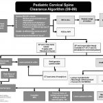
Algorithm For Clearing the Pediatric Cervical Spine

I previously wrote about a straightforward way to clear the cervical spine in children. Click here to see the article. Alfred I. DuPont Children’s Hospital has condensed their clearance technique into a relatively simple algorithm that can be used in conjunction with my previous tips.

Some notes on this algorithm:

  • Can be performed only by attending physicians or a trauma resident in consultation with the attending trauma surgeon
  • Clinical clearance alone may be carried out in select cases
  • If radiographs are required, cross-table lateral, anterior/posterior, and odontoid views should be obtained (age 8 and above, non-intubated)
  • Flexion / extension views should only be ordered in consultation with neurosurgery

Download a print version of the protocol here

Related post: How Do I Clear The Pediatric Cervical Spine?

Image and protocol courtesy of the Alfred I DuPont Children’s Hospital

Pneumothorax in Children

Last week I wrote about pneumomediastinum in children (click here to read). This week I’ll talk about a somewhat more common problem: pneumothorax. This condition is far more mysterious than in adults.

Sports related pneumothorax rarely occurs without rib fractures, which are themselves uncommon in children. The usual mechanism is barotrauma, most likely from an impact while the glottis is closed. The typical presentation is that of pleuritic chest pain, which may be followed by dyspnea. Focal chest wall tenderness is typically absent. Teenagers tend to engage in more strenuous activity and are more likely to actually sustain a rib fracture, so they may have focal tenderness over the fracture site.

Spontaneous pneumothorax in children is also uncommon. However, it is a very different entity. It may be related to blebs in the lungs, and may be more common in children who were born prematurely. The recurrence rate after successful treatment is approximately 50% (in small series). Recurrence is not predictable by looking for blebs on chest CT. The recurrence rate is significantly lower after VATS.

Bottom line: A child who complains of pleuritic chest pain, and especially dyspnea, should undergo a simple PA chest xray. If a pneumothorax is present, consider the following:

  • Insert a small chest tube or catheter if needed, the smaller the better. (I’m stilling looking for the answer to the question of how big a pneumothorax is big enough)
  • Don’t use high inspired oxygen; it doesn’t work. (Read my older blogs from 2010 – this one and this one, too)
  • Don’t get a chest CT for either the initial pneumothorax or any recurrences (too much radiation, too little utility)
  • If this is a spontaneous pneumothorax, caution the parents on the possibility of recurrence
  • If a spontaneous pneumothorax does recur, consult a pediatric surgeon to consider VATS pleurodesis
  • When can the child return to sports? There is absolutely no good literature. I recommend the usual time it takes most soft tissues to return to full tensile strength after injury (6 weeks).

References:

  • Management of primary spontaneous pneumothorax in children. Clin Pediatr, online ahead of print, April 11, 2011.
  • Sports-related pneumothorax in children. Pediatric Emergency Care 21(4): 259-260, 2005.

Related posts:

Thanks to Chris Chow MD for finding some of the literature for this post.

How Significant is Pneumomediastinum in Children?

Pneumomediastinum seen on chest x-ray after blunt trauma always attracts attention. Possible sources may be related to very serious injuries to the aerodigestive tract. When seen in children, it causes considerable anxiety, which usually results in a very detailed workup and lots of imaging.

Children’s Hospital of Boston looked at the National Trauma Data Bank, as well as 19 years worth of their own records to see whether all the attention is justified. They found 193 patients in the NTDB that met their criteria, and most were in their late teens and had other significant injuries. Of the 17 with isolated pneumomediastinum, none had any other significant injury.

When reviewing their own patient records, they found 18 with pneumomediastinum, and all but one was seen on plain chest x-ray. Most were transferred to the hospital from referring centers, and had been involved in sports-related mechanisms. Half had undergone studies in addition to a chest x-ray before transfer. All were discharged home without any surgical interventions.

Bottom line: Pneumomediastinum is rare in children, even older ones. If associated with significant aerodigestive injuries, it was never an isolated occurrence. Other signs or symptoms were present. Pediatric patients presenting with an isolated pneumomediastinum can be safely observed, using chest x-ray and physical examination alone. More sophisticated studies (CT, barium studies) are not indicated.

Reference: Clinical outcomes and diagnostic imaging of pediatric patients with pneumomediastinum secondary to blunt trauma  to the chest. J Trauma, epub, 2011.

Trauma 20 Years Ago: Blunt Aortic Injury in Children

We always worry about the aorta after high-energy blunt trauma in adults. Should we be doing the same in kids? After all, they are very elastic and for the most part they are tough to break.

A 13 year review was undertaken by the CV surgeons at Harborview twenty years ago which tried to answer this question. They looked at medical examiner records of all pediatric deaths (16 or younger) and identified the ones with traumatic aortic injury. They found only 12 deaths (2.1%), and somehow they also tracked one survivor (from ME data???). The age range was 3-15, with a mean of 12 (which means that the majority were in the older age group).

Six children were pedestrian struck, 5 were involved in car crashes, and two were on motorized bikes or ATVs. None of the children in car crashes were restrained and two were ejected. Four of the five were traveling > 55mph. All had other serious injuries, including abdominal and orthopedic.

It’s tough to draw any meaningful conclusions from this paper due to the small numbers, the retrospective design, and the lack of a denominator. The only thing it does tell us is that aortic injury is bad, and that kids should not get hit by cars and should wear their seat belts. The mean age suggests that it involves primarily older children. But we kind of knew all that already.

What it does not help with is figuring out at what age we need to start thinking about imaging the aorta with CT scan. I’ll be digging into that a little more this week.

Reference: Eddy et al. The epidemiology of traumatic rupture of the aorta in children: a 13 year review. J Trauma 30(8): 989-992, 1990.