Tag Archives: hemorrhage

What Is: Life Threatening Bleeding In The Anticoagulated Patient – Part 1

We are moving into a world where more and more people are taking anticoagulants. And as the number of possible anticoagulant drugs increases, the prospect of reversing them becomes more complicated. Just as the price of those medications continues to climb, so does the price of the agents used to reverse them.

The decision to reverse an anticoagulant, the speed of reversal, and choice of reversal agent all depend on an assessment of the severity of bleeding. Some reversal drugs such as prothrombin complex concentrate (PCC) act almost immediately but are expensive. Others take time, but are cheaper such as plasma and vitamin K.

Unfortunately, trying to come to a consensus on what constitutes life-threatening bleeding is very difficult. Over the years, numerous studies have been done, with almost as many definitions of bleeding. As you know, I am generally against reinventing the wheel. Borrowing someone else’s excellent work saves a lot of time and anguish.

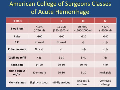
But when it comes to defining dangerous bleeding, we are faced with so many definitions, it just begs for a “unifying theory.” I’ll show you some of the more commonly used definitions below. At the very bottom, I’ve included a link to a very comprehensive list of definitions that have been used.

First, there’s TIMI (Thrombolysis in Myocardial Infarction trial) and GUSTO (Global Use of Strategies to Open Occluded Arteries trial). (Please remember how much I dislike cute acronyms.)

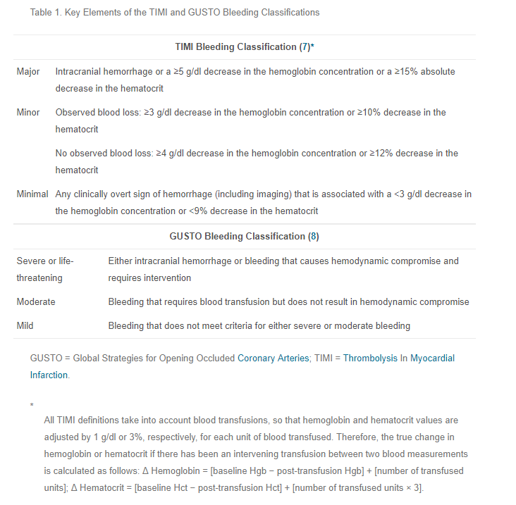
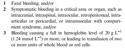
The following consolidated definition was published in the Journal of Thrombosis and Haemostsis way back in 2005:

If you want to look at a more comprehensive list of a lot of definitions, download the document from the link below.

So how do we make sense of all this? As trauma professionals and clinicians on the front line of anticoagulant reversal, we need a simple definition. I’ve recently looked over as many definitions as I could lay my hands on. 

In my next post, I’ll propose a simplified set of definitions. And I’ll be very interested in your input and comments. They will ultimately end up as a definition that we will use at my own trauma center. And maybe yours.

Related post:

Reference:

  • Definition of major bleeding in clinical investigations of antihemostatic medicinal products in non-surgical patients. Journal of Thrombosis and Haemostasis, 3: 692–694, 2005.

How To Remember Those “Classes of Hemorrhage”

The Advanced Trauma Life Support course lists “classes of hemorrhage”, and various other sources list a similar classification for shock. I’ve not been able to pinpoint where these concepts came from, exactly. But I am sure of one thing: you will be tested on it at some point in your lifetime.

Here’s the table used by the ATLS course:

classes_of_shock

The question you will always be asked is:

What class of hemorrhage (or what % of blood volume loss) is the first to demonstrate systolic hypotension?

This is important because prehospital providers and those in the ED typically rely on systolic blood pressure to figure out if their patient is in trouble.

The answer is Class III, or 30-40%. But how do you remember the damn percentages?

multiscore-maxi1

It’s easy! The numbers are all tennis scores. Here’s how to remember them:

Class I up to 15% Love – 15
Class II 15-30% 15 – 30
Class III 30-40 30 – 40
Class IV >40% Game (almost) over!

Bottom line: Never miss that question again!

Can Prehospital Providers Accurately Estimate Blood Loss? Part 2

I’ve previously written about the difficulties estimating how much blood is on the ground at the trauma scene. In general, EMS providers underestimated blood loss 87% of the time. The experience level of the medic was of no help, and the accuracy actually got worse with larger amounts of blood lost!

A group in Hong Kong developed a color coded chart (nomogram) to assist with estimation of blood loss at the scene. It translated the area of blood on a non-absorbent surface to the volume lost. A convenience study was designed to judge the accuracy that  could be achieved using the nomogram. Sixty one providers were selected, and estimated the size of four pools of blood, both before and after a 2 minute training session on the nomogram.

Here’s what it looks like:

Note the areas across the bottom. In addition to colored square areas, the orange block is a quick estimate of the size of a piece of paper (A4 size since they’re in Hong Kong!)

Here are the factoids:

  • The 61 subjects had an average of 3 years of experience
  • Four scenarios were presented to each: 180ml, 470ml, 940ml, and 1550ml. These did not correspond exactly to any of the color blocks.
  • Before nomogram use, underestimation of blood loss increased as the pool of blood was larger, similar to the previous study
  • There was a significant increase in accuracy for all 4 scenarios using the nomogram, and underestimation was significantly better for all but the 940ml group
  • Median percentage of error was 43% before nomogram training, vs only 23% after. This was highly significant.

Bottom line: This is a really cool idea, and can make estimation of field blood loss more accurate. All the medic needs to do is know the length of their shoe and the width of their hand in cm. They can then estimate the length and width of the pool of blood and refer to the chart . Extrapolation between colors is very simple, just look at the line. The only drawback I can see occurs when the blood is on an irregular or more absorbent surface (grass, inside of a car). 

Related posts:

Reference:  Improvement of blood loss volume estimation by paramedics using a pictorial nomogram: a developmental study. Injury article in press Oct 2017.

How To Remember Those “Classes of Hemorrhage”

The Advanced Trauma Life Support course lists “classes of hemorrhage”, and various other sources list a similar classification for shock. I’ve not been able to pinpoint where these concepts came from, exactly. But I am sure of one thing: you will be tested on it at some point in your lifetime.

Here’s the table used by the ATLS course:

classes_of_shock

The question you will always be asked is:

What class of hemorrhage (or what % of blood volume loss) is the first to demonstrate systolic hypotension?

This is important because prehospital providers and those in the ED typically rely on systolic blood pressure to figure out if their patient is in trouble.

The answer is Class III, or 30-40%. But how do you remember the damn percentages?

multiscore-maxi1

It’s easy! The numbers are all tennis scores. Here’s how to remember them:

Class I up to 15% Love – 15
Class II 15-30% 15 – 30
Class III 30-40 30 – 40
Class IV >40% Game (almost) over!

Bottom line: Never miss that question again!

Delayed Intracranial Hemorrhage In Patients On Anticoagulants

A sizable portion of our population is taking one type of anticoagulant or another. Heck, even golf star Arnold Palmer and comedian Kevin Nealon are on Xarelto! Any trauma professional, and anyone who reads the package insert, knows that there is an increased risk of bleeding if they are injured while taking these drugs, whether it be warfarin or the new, novel anticoagulants.

But does the risk stop soon after injury? That is the presumption at many hospitals that initially treat these patients. They are seen in the ED, examined, scanned, and sent home if nothing is found. Is this a safe practice?

I have personally seen a patient who had an initially clean CT present within 12 hours after ED discharge with a catastrophic bleed and die. Yes, this is anecdotal, but I have talked to other trauma professionals with similar experiences. If this were just a minor complication, no big deal. But they died. Big problem for everyone involved.

So what does the literature say? Unfortunately, it consists of a collection of relatively small studies. Here are the collected factoids that I can glean from them:

  • Most are retrospective, observational studies 
  • Most are from a single hospital, which may miss readmissions to other facilities in the area
  • The delayed bleeding rate is about 0.5% to 1%
  • Some papers recommended discharging patients with a normal head CT and giving them instructions to return if new symptoms develop (this is what happened with my patient; what if they live alone or in a care center where these may not be recognized?!)
  • A few papers did identify patients needing neurosurgical intervention or who died
  • Immediate bleeds were more common with antiplatelet agents, delayed bleeds were more common with warfarin
  • I could find nothing that looked at this problem in patients taking novel anticoagulants like Pradaxa or Xarelto

Bottom line: The literature provides little guidance at this point. A good multi-institutional trial is needed to generate the numbers to tell us what to do. While we get around to this, I recommend that a selective brief observation (12 hrs) protocol be adopted. This protocol recognizes that subclinical bleeding may be present on initial presentation, and that a little more time is needed for it to declare itself.

Here is a link to our protocol. If the initial head CT is negative and the INR is less than 2.5, we will only discharge the patient if all of these criteria are true:

  • Age < 65
  • No skull fx
  • No new focal neurologic deficits
  • No soft tissue injury visible on CT (hematoma, laceration)
  • GCS = 15
  • No persistent vomiting
  • Brief TBI screen passed (Short Blessed Test, link here)

Most do not pass all of these, usually failing the age criterion. They are admitted for observation and neurologic monitoring for 12 hours, at which time the head CT is repeated. If it is still normal, then they can go home.

And although this protocol was designed with warfarin in mind, we apply it to patients taking novel anticoagulants like Pradaxa and Xarelto as well. We’ve had no epic fails yet, but I keep my fingers crossed!

Related posts:

References:

  • Management of minor head injury in patients receiving oral anticoagulant therapy: a prospective study of a 24-hour observation protocol. Ann Emerg Med 59(6):451-455, 2012.
  • Immediate and delayed traumatic intracranial hemorrhage in patients with head trauma and preinjury warfarin or clopidogrel use. Ann Emerg Med 59(6):460-468, 2012.
  • Delayed intracranial hemorrhage after blunt trauma: are patients on preinjury anticoagulants and prescription antiplatelet agents at risk? J Trauma 71(6):1600-1604, 2011.
  • Low risk of late intracranial complications in mild traumatic brain injury patients using oral anticoagulation after an initial normal brain computed tomography scan: education instead of hospitalization. Eur J Neurol 21(7):1021-1025, 2014.
  • Can anticoagulated patients be discharged home safely from the emergency department after minor head injury? J Emerg Med 46(3):410-417, 2014.
  • Patients with blunt head trauma on anticoagulation and antiplatelet medications: can they be safely discharged after a normal initial cranial computed tomography scan? Am Surg 80(6):610-613, 2014.