Tag Archives: hemorrhage

Interesting Concept: The Abdominal Aortic & Junctional Tourniquet

Tourniquets for extremity bleeding are definitely back in vogue. Our military experience over the past 20 years has shown us what a life saver this simple tool can be. It’s now carried by many prehospital trauma professionals for use in the civilian population. But what about bleeding from the nether regions? You know what I’m talking about, the so-called junctional zones. Those are the areas that are too proximal (or too dangerous) to put on a tourniquet, like the groin, perineum, axilla, and neck.

Traditionally, junctional zone injury could only be treated in the field with direct pressure, clamps, or in some cases a balloon (think 30Fr Foley catheter inserted and blown up as large as possible, see link below). In the old days, we could try blowing up the MAST trousers to try to get a little control, but those are getting hard to find. 

An Alabama company (Compression Works) developed a very novel concept to try to help, the Abdominal Aortic and Junctional Tourniquet (AAJT). Think of it as a pelvic compression device that you purposely apply too high.

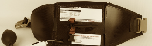
Note the cool warning sticker at the bottom of the device!

The developers performed a small trial on 16 volunteer soldiers after doing a preliminary test on themselves (!). The device was placed around the abdomen, above the pelvis, and inflated to a maximum of 250 torr. Here are the factoids:

  • All subjects tolerated the device, and no complications occurred
  • Flow through the common femoral artery stopped in 15 of the 16 subjects
  • The subject in whom it did not work exceeded the BMI and abdominal girth parameters of the device
  • Average pain score after application was 6-7 (i.e. hurts like hell!)

Here’s a list of the criteria that preclude use of this device:

Bottom line: This would seem to be a very useful device for controlling hemorrhage from pesky areas below the waist.

BUT! Realistically, it will enjoy only limited use in the civilian population for now. Take a closer look at the exclusion criteria above. Half of the population is ineligible right off the bat (women). And among civilians, more than a third are obese in the US. Toss in a smattering of the other criteria, and the unlikelihood of penetrating trauma to that area in civilians, it won’t make financial sense for your average prehospital agency to carry it. Maybe in high violence urban areas, but not anywhere else.

The company has received approval for use in pelvic and axillary hemorrhage control, so we’ll see how it works when more and larger studies are released (on more and larger people). 

Related post:

Reference: The evaluation of an abdominal aortic tourniquet for the control of pelvic and lower abdominal hemorrhage. Military Med 178(11):1196-1201, 2013.

Thanks to David Beversluis for bringing this product to my attention. I have no financial interest in Compression Works.

Best Of How To: Stop Scalp Bleeding

Bleeding from scalp wounds may seem like a trivial problem, but I have personally seen someone die from unrecognized hemorrhage over time from one. All too often, these are covered up with a crude dressing when the patient arrives in the ED and is not looked at for some time.

Here are some tips to stop scalp bleeding:

  • Use direct pressure. This seems obvious but is frequently done incorrectly. Direct pressure involves a small diameter piece of gauze (stack of 2x2s or double folded 4×4) and only one or two fingers. Larger dressings or the palm of the hand do not provide enough pressure to stop all the bleeding. Direct pressure for 5 minutes (no peeking) will stop all bleeding that doesn’t need more advanced techniques.
  • Inject local anesthetic with epinephrine. This increases vasoconstriction and helps the direct pressure work even better. Be cautious if there is a large skin flap that does not have a nice pink color. Degloved skin has been crushed and small vessel vascular injury has occurred. Further reducing blood flow with epinephrine may kill the skin flap in this type of injury.
  • Apply Raney clips. Neurosurgeons use these to stop scalp bleeding during brain procedures. Caution! Only apply to unconscious patients, and only to the scalp (not face)! These hurt!
    Raney clips
  • Oversew the scalp. Use a large silk or nylon suture and insert a large running stitch to close the wound. This will stop all bleeding from the skin edges. However, any arterial bleeders underneath will continue to be a problem.
  • Ligate individual bleeders. Use a small absorbable suture and attack each small arterial bleeder with a figure of 8 stitch. Don’t suture large chunks of tissue; make sure that you are attacking just the artery and not any adjacent nerves.

AAST 2011: The Initial Hematocrit Matters

Traditional teaching is that we bleed whole blood, and it takes time to pull volume out of the interstitial space to replace it. Therefore, the initial hematocrit should be normal when a fresh, bleeding trauma patient rolls through the doors.

An observation I have made over the years is that this is not necessarily so. A few patients have low initial hemoglobin or hematocrit readings, and they tend to be bleeding briskly from somewhere. A paper to be presented at next week’s AAST meeting in Chicago shows just that.

The authors retrospectively reviewed 198 trauma patients requiring emergency surgery at a Level I trauma center. Patients with lower initial hematocrits tended to have lower systolic blood pressure, lower GCS, lose more blood, and require infusion of more blood products during surgery. They also had a higher ISS and mortality. The biggest jump in these indicators occurred when the Hct dropped below 37.

Bottom line: A low hematocrit on the first blood drawn during trauma resuscitation is more helpful that previously thought. Be sure to check those lab values early, and if the hematocrit value is in the mid-30s or lower, start looking for significant sources of bleeding.

Reference: The initial hematocrit matters in trauma: a paradigm shift? AAST 2011 Annual Meeting, Paper 38.

Bleeding and Pelvic Fractures

Arterial bleeding from a pelvic fracture is more common than previously thought. The doctor books used to say that 10% of bleeding was arterial and 90% was venous, so angiographic techniques were seldom used unless there was clinical evidence of blood loss. 

It looks like arterial bleeding occurs more frequently than we think. Here are tips that help you identify patients at risk:

  • What type of mechanism caused the fracture? Anterior-posterior compression and vertical shear are the most common.
  • Are the vital signs stable? If not, rule out the other four likely sources first (chest, abdomen, multiple extremity fractures, external). Then blame the pelvis.
  • Is the fracture open? Arterial bleeding is very likely.
  • How old is the patient? Elderly patients are more likely to have arterial bleeding, especially from gluteal artery branches.
  • What part of the pelvis is broken? If major sacral fractures, SI joint disruption or separation of the symphysis is present, think arterial bleeding.
  • Are there CT abnormalities? A vascular blush or large hematoma indicates significant bleeding.

The most common bleeding sites are the gluteal and pudendal arteries. The gluteal is in proximity to the SI joint, so this can be torn if the SI joint is damaged or the sacrum is fractured. The pudendal can be injured with ramus fractures, especially when the symphysis is widened.

If the patient can be reasonably stabilized, then a trip to interventional radiology is mandatory. Operative management is not very successful, so patients with blood pressure lability or controllable hypotension should go to IR. All active bleeding and arterial cutoffs should be embolized thoroughly.

Images: On the left is the portable plain image of a vertical shear pelvic fracture. The arrows on the right point to two areas of vascular blush.

Pelvic arterial bleeding

How Accurate is EMS at Estimating Blood Loss in the Field?

EMS providers are the trauma professional’s eyes and ears when providing transportation from the scene of an accident. We rely on their assessment of the mechanism of injury and the amount of blood lost. We tend to believe in the accuracy of those assessments.

A study was carried out that tested EMS personnel on their ability to accurately estimate specific amounts of blood that were left at a simulated accident scene. The blood volumes tested were 500cc, 1000cc, 1500cc and 2100cc. A total of 92 professionals participated, and there was an even split into basic EMTs (34%), intermediate/critical care EMTs (33%) and paramedics (31%). Experience levels were as follows: 0-5 years 43%, 6-10 years 30%, >10 years 31%.

The results were as follows:

  • 87% underestimated the quantity of blood
  • 9% overestimated
  • 4% guessed the exact amount
  • Experience or credentialing level did not matter

Only 8% of the subjects were within 20% of the actual volume, and an additional 19% were within 50%. In general, most medics underestimated the amount of blood lost, and their guesses were worse with higher volumes. The median guess for the 2100cc loss group was only 700cc!

EMS Blood Loss Estimates

The bottom line: Visual estimates of blood loss are extremely inaccurate, and are most likely  underestimates. Physicians in the ED should rely on exam and physiology to help determine the amount of blood loss. For safe measure, multiply the reported blood loss of the EMT or paramedic by 2 or 3 to get a realistic number.

Reference: Patton et al. Accuracy of Estimation of External Blood Loss by EMS Personnel. J Trauma 50(5), 914, 2001.