Tag Archives: CT

If A Tree Falls In A Forest…

Time for a little philosophy today. There seem to be two camps in the world of initial diagnostic testing for trauma: selective scanning vs scan everything. I admit that I am one of the former. Yes, the more tests you do, the more things you will find. Some will be red herrings. Some may be true positives, but are they important? Here’s the key question:

“If a tree falls in a forest and no one is around, does it make a sound?”

There is a clinical corollary to this question in the field of trauma:

“If an injury exists but no one diagnoses it, does it make a difference (if there would be no change in treatment)?”

Here’s an example. On occasion, my colleagues want to order diagnostic studies that won’t make any clinical difference, in my opinion. A prime example is getting a chest CT after a simple blunt assault. A plain chest xray is routine, and if injuries are seen or the physical exam points to certain diagnoses, appropriate interventions should be taken. But adding a chest CT does not help. Nothing more than the usual pain management, pulmonary toilet, and an occasional chest tube will be needed, and those can be determined without the CT.

Trauma professionals need to realize that we don’t need to know absolutely every diagnosis that a patient has. Ones that need no treatment are of academic interest only, and can lead to accidental injury if we look for them too hard (radiation exposure, contrast reaction, extravasation into soft tissues to name a few). This is how we get started on the path to “defensive medicine.”

Bottom line: Think hard about every test you order. Consider what you are looking for, what you might find, and if it will change your management in any way. If it could, go ahead. But always consider the benefits versus the potential risks, or what I call the “juice to squeeze ratio.”

Tomorrow I’ll look at some of the “scan all” vs “scan selectively” literature. Which camp are you in?

References:

  • George Berkeley, A Treatise Concerning the Principles of Human Knowledge, 1734, section 45.
  • paraphrased by William Fossett, Natural States, 1754.

Identifying Bowel and Mesenteric Injury by CT

CT scan is an invaluable tool for evaluating blunt abdominal trauma. Although it is very good at detecting solid organ injury, it is not so great with intestinal and mesenteric injuries. Older studies have suggested that CT can detect mesenteric injuries if done right, but a newly published study has shown good accuracy with a few imaging tweaks.

A Taiwanese study looked at a series of prospectively studied victims of blunt abdominal trauma. Patients with abdominal pain or a positive FAST were entrolled (total 106). IV contrast was given, and scans during the arterial, portal, and equilibrium contrast phases were performed using a multidetector scanner. Images were read in a blinded fashion.

A total of 13 of 23 patients who underwent laparotomy were found to have a bowel or mesenteric injury. Five had bowel injury, 4 had mesenteric hemorrhage, and 4 had both. Mesenteric contrast extravasation was seen in 7 patients, and this correlated with mesenteric bleeding at laparotomy.

The authors found that the following signs on CT scan indicated injury:

  • Full or partial thickness change in bowel wall appearance
  • Increased mesenteric density
  • Free fluid without solid organ injury

Bottom line: This study shows that CT scan can detect bowel and mesenteric injury reliably if you scan the patient 3 times! This seems like over-radiation and overkill. A more intelligent way to approach this would be to perform a normal trauma abdominal scan. If a suspicious area of mesenteric or bowel thickening is seen, then a limited rescan through the affected area only for equilibrium phase images may be warranted. If actual contrast extrvasation is seen, no further scanning is needed. A quick trip to the OR is in order.

Reference: Contrast-enhanced multiphasic computed tomography for identifying life-threatening mesenteric hemorrhage and transmural bowel injuries. J Trauma 71(3):543-548, 2011.

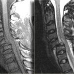
Clearing The Cervical Spine With MRI

If you follow the trauma literature, clearance of the cervical spine in obtunded patients is confusing at best. Although there is some literature out there that suggests that a good cervical CT alone is adequate, I’m not a believer. I’ve seen a case where the radiologist called the scan normal and a good spine surgeon called an injury and was right. So I’m reluctant to use CT alone because the skills of radiologists vary widely. I might be able to believe a dedicated neuroradiologist, but you can’t guarantee one will be reading your patient’s images.

So I fall back on the routine of clearing the bones with a CT scan, and the ligaments with something else. That something else could be a clinical exam (not available in the obtunded patient), flexion-extension images under fluoroscopy (makes a lot of people nervous), keeping the patient in a collar for weeks (skin breakdown), or an MRI. The problem is that there is little guidance in the literature regarding how good MRI is or the best way to use it.

A recent paper in the Journal of Trauma retrospectively looked at 512 out of 17,000 patients (!) seen over 5 years at one trauma center who had both CT and MRI of the c-spine. They wanted to determine if MRI was of any value in cervical spine clearance. Only 150 met the inclusion criteria (GCS<13, no obvious neuro deficit, normal CT). Half of the MRIs were normal. Of the abnormal ones, 81% showed a ligamentous or soft tissue injury. None were deemed unstable and no specific management was needed for any of the abnormal scans.

The authors interpreted their data as showing that MRI provided no additional useful information. However, numbers were (very) small, so the likelihood of them seeing someone with an unstable ligamentous injury was low. Could it be that they showed that MRI detected stable injuries well, and that they could essentially remove the collar based on that?

Bottom line: We still don’t know how to use MRI for clearance. My bias (no good data I can find) is that it is good in suggesting ligamentous injury via nearby edema. If this injury involves only one set of ligaments, it is very likely a stable one and the collar can be removed. If it involves several groups of ligaments, that is probably not the case. And how soon do we have to get the MRI after injury? Some have suggested that 72 hours is the ideal window because edema decreases afterwards. Sounds reasonable, but I can’t find a shred of evidence in the literature. For now, I’ll get an MRI within 72 hours and if it is abnormal, pass the buck to my neurosurgical colleagues so they can gnash their teeth, too.

I would be very happy if someone can help me out and point me towards some good literature on this topic!

Reference: The value of cervical magnetic resonance imaging in the evaluation of the obtunded or comatose patient with cervical trauma, no other abnormal neurological findings, and a normal cervical computed tomography. J Trauma 72(3):699-702, 2012.

CT Scan Images Simplified

Ever wonder what is going on when you drag your mouse across a CT image, or when you change the “window” settings of an image from lung to abdomen? It all has to do with the way CT generated xray information is displayed, and how your eyes and brain perceive it.

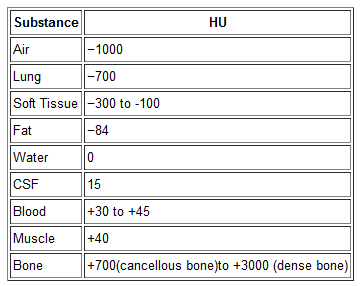
Let’s get down to basics. The first thing needed is to understand the concept of radiodensity. The CT scanner uses a set of software algorithms to determine the amount of x-radiation absorbed by every element in a plane of tissue. Each of these elements is represented by a pixel on the video display, and the density (amount of x-radiation absorbed) is measured in Hounsfield units. This scale was developed by Sir Godfrey Hounsfield, who set the radiodensity of water at 0, and air at -1000. The scale extends in the positive direction to about +4000, which represents very dense metals. See the table for the density of common substances on CT.

When you view a CT scan on a video display, two important numbers are displayed on screen. The first is the window width (W), which describes the range of Hounsfield units displayed. The maximum window width possible is usually about 2000, but our eyes are not capable of seeing this many shades. Actually, we can really only distinguish about 16 shades of gray. So the window width is divided by 16, and each group of Hounsfield values is converted to one of 16 shades of gray. The lowest Hounsfield numbers in the window range are shown as black, and the highest are white.

The second important number is the window level (L). This is the Hounsfield number in the center of the window width. So let’s look at some typical examples of W/L settings.

The abdomen contains mostly soft tissue, which is just a little denser than water. So most of the abdominal contents have Hounsfield values from 0 to 100 or so. A typical abdominal scan W/L setting is 350/50. This means that a total range of 350 different densities are displayed, centered on a density of 50 Hounsfield units ( range is -125 to 225 HU). Each difference of 22 HU will show up as a different shade of gray. So this narrow window allows us to distinguish relatively subtle differences in density.

The chest cavities are primarily air-filled, and the lungs are very low density. So it makes sense that a typical lung W/L setting is 1500/-500. The window ranges from -1250 to +250 HU, and a wider range of 94 HU represents one shade of gray. This is typical of body regions with a wider range of densities.

Finally, bone windows are usually 2000/250. This window is centered above the usual tissue densities, and is very wide so that it shows a wide range of densities in only 16 shades of gray. Thus, the contrast appears very low.

On most displays, the window width increases as you drag the mouse to the right. This increases the range of densities in a shade of gray, thus decreasing the overall amount of contrast in the image. Dragging the mouse down decreases the window level, moving it toward the air end of the spectrum. This allows you to center your window on the type of tissue you are interested in viewing and adjust your ability to distinguish objects with a lot or only a little contrast (see table above).

I apologize to my radiology colleagues in advance for this simplistic explanation. Trauma professionals have minimal exposure (pun intended) to the physics and details of radiographic imaging. We are much more interested in effectively using this technology to save our patients’ lives.

Identifying Bowel and Mesenteric Injury by CT

CT scan is an invaluable tool for evaluating blunt abdominal trauma. Although it is very good at detecting solid organ injury, it is not so great with intestinal and mesenteric injuries. Older studies have suggested that CT can detect mesenteric injuries if done right, but a newly published study has shown good accuracy with a few imaging tweaks.

A Taiwanese study looked at a series of prospectively studied victims of blunt abdominal trauma. Patients with abdominal pain or a positive FAST were entrolled (total 106). IV contrast was given, and scans during the arterial, portal, and equilibrium contrast phases were performed using a multidetector scanner. Images were read in a blinded fashion.

A total of 13 of 23 patients who underwent laparotomy were found to have a bowel or mesenteric injury. Five had bowel injury, 4 had mesenteric hemorrhage, and 4 had both. Mesenteric contrast extravasation was seen in 7 patients, and this correlated with mesenteric bleeding at laparotomy.

The authors found that the following signs on CT scan indicated injury:

  • Full or partial thickness change in bowel wall appearance
  • Increased mesenteric density
  • Free fluid without solid organ injury

Bottom line: This study shows that CT scan can detect bowel and mesenteric injury reliably if you scan the patient 3 times! This seems like over-radiation and overkill. A more intelligent way to approach this would be to perform a normal trauma abdominal scan. If a suspicious area of mesenteric or bowel thickening is seen, then a limited rescan through the affected area only for equilibrium phase images may be warranted. If actual contrast extrvasation is seen, no further scanning is needed. A quick trip to the OR is in order.

Reference: Contrast-enhanced multiphasic computed tomography for identifying life-threatening mesenteric hemorrhage and transmural bowel injuries. J Trauma 71(3):543-548, 2011.