Tag Archives: CT

Torso Trauma CT (Nearly) ALWAYS Requires Contrast

Most stable patients with blunt trauma undergo CT scanning these days. Hopefully, it’s done thoughtfully to optimize the risk/benefit ratio using a well-designed imaging protocol. The majority of these torso imaging protocols call for the use of IV contrast. But as I’ve written before, this can pose risks, especially to the elderly and others who have some degree of renal impairment.

Unfortunately, I occasionally encounter scans done at other hospitals that omit the use of contrast. This usually hinders diagnosis significantly. And it’s usually not clear why this happened, so let’s think about it a bit.

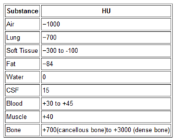
The use of contrast in CT is designed to show blood, or things that are filled with lots of blood. Specifically, a great deal of detail about the blood vessels and solid organs is displayed.

Let’s break it down by type of scan:

  • Chest – we are really only interested in the aorta. The only way to reliably demonstrate an aortic injury is by using contrast. And this is one of those injuries that, if you miss it, the patient is very likely to die from it. Therefore, if you are ordering a chest CT properly, you must add contrast.
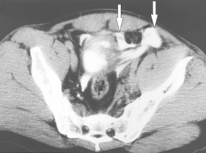
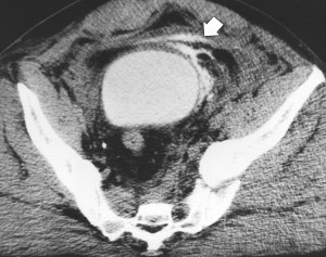
  • Abdomen/pelvis – generally, we are looking for solid organ injury, potential mesenteric injuries, and extravasation of blood from organs or soft tissue. Once again, the only way to really see any of these is with contrast enhancement.
  • Vascular – CT is replacing conventional angiography for the investigation of vascular injury in many cases. Obviously, this study is worthless without the contrast.

Bottom line: Pretty much any CT of the chest, blood vessels, or abdomen/pelvis must have IV contrast injected for accurate diagnosis. But what if your patient is old, or is known to have some degree of renal impairment? First, decide if you can wait until a point of care or standard creatinine measurement is done. If you can, use the result to do your own risk/benefit calculation. Is the injury you are worried about potentially life-threatening AND reasonably likely? Are there other less harmful ways to detect it? Then use them. And if you really do need the study in a patient with renal dysfunction, give the contrast, monitor the serum creatinine regularly, and do what you can to optimize and protect their renal function over the next several days.

CT Cystography For Bladder Trauma

Bladder injury after blunt trauma is relatively uncommon, but needs to be identified promptly. Nearly every patient (97%+) with a bladder injury will have hematuria that is visible to the naked eye. This should prompt the trauma professional to obtain a CT of the abdomen/pelvis and a CT cystogram.

The CT of the abdomen and pelvis will identify any renal or ureteral (extremely rare!) source for the hematuria. The CT cystogram will demonstrate a bladder injury, but only if done properly!

During most trauma CT scanning of the abdomen and pelvis, the bladder is allowed to passively fill, either by having no urinary catheter and having the patient hold it, or by clamping the catheter if it is present. Unfortunately, this does not provide enough pressure to demonstrate small intraperitoneal bladder injuries and most extraperitoneal injuries.

The proper technique involves infusing contrast into the bladder through a urinary catheter. At least 350cc of dilute contrast solution must be instilled for proper distension and accurate diagnosis. This can be done prior to the abdominal scan. Once the initial scan has been obtained, the bladder must be emptied and a focused scan of just the bladder should be performed (post-void images). Several papers have shown that this technique is as accurate as conventional retrograde cystography, with 100% sensitivity and specificity for intraperitoneal ruptures. The sensitivity for extraperitoneal injury was slightly less at 93%.

Bottom line: Gross hematuria equals CT of the abdomen/pelvis and a proper CT cystogram, as described above. Don’t try to cheat and passively fill the bladder. You will miss about half of these injuries!

Related posts:

Reference: CT cystography with multiplanar reformation for suspected bladder rupture: experience in 234 cases. Am J Roentgenol 187(5):1296-302, 2006.

Intraperitoneal bladder rupture

Extraperitoneal bladder injury

CT Scan Image Settings Simplified

Ever wonder what is going on when you drag your mouse across a CT image, or when you change the “window” settings of an image from lung to abdomen? It all has to do with the way CT generated xray information is displayed, and how your eyes and brain perceive it.

Let’s get down to basics. The first thing needed is to understand the concept of radiodensity. The CT scanner uses a set of software algorithms to determine the amount of x-radiation absorbed by every element in a plane of tissue. Each of these elements is represented by a pixel on the video display, and the density (amount of x-radiation absorbed) is measured in Hounsfield units. This scale was developed by Sir Godfrey Hounsfield, who set the radiodensity of water at 0, and air at -1000. The scale extends in the positive direction to about +4000, which represents very dense metals. See the table for the density of common substances on CT.

When you view a CT scan on a video display, two important numbers are displayed on screen. The first is the window width (W), which describes the range of Hounsfield units displayed. The maximum window width possible is usually about 2000, but our eyes are not capable of seeing this many shades. Actually, we can really only distinguish about 16 shades of gray. So the window width is divided by 16, and each group of Hounsfield values is converted to one of 16 shades of gray. The lowest Hounsfield numbers in the window range are shown as black, and the highest are white.

The second important number is the window level (L). This is the Hounsfield number in the center of the window width. So let’s look at some typical examples of W/L settings.

The abdomen contains mostly soft tissue, which is just a little denser than water. So most of the abdominal contents have Hounsfield values from 0 to 100 or so. A typical abdominal scan W/L setting is 350/50. This means that a total range of 350 different densities are displayed, centered on a density of 50 Hounsfield units ( range is -125 to 225 HU). Each difference of 22 HU will show up as a different shade of gray. So this narrow window allows us to distinguish relatively subtle differences in density.

The chest cavities are primarily air-filled, and the lungs are very low density. So it makes sense that a typical lung W/L setting is 1500/-500. The window ranges from -1250 to +250 HU, and a wider range of 94 HU represents one shade of gray. This is typical of body regions with a wider range of densities.

Finally, bone windows are usually 2000/250. This window is centered above the usual tissue densities, and is very wide so that it shows a wide range of densities in only 16 shades of gray. Thus, the contrast appears very low.

On most displays, the window width increases as you drag the mouse to the right. This increases the range of densities in a shade of gray, thus decreasing the overall amount of contrast in the image. Dragging the mouse down decreases the window level, moving it toward the air end of the spectrum. This allows you to center your window on the type of tissue you are interested in viewing and adjust your ability to distinguish objects with a lot or only a little contrast (see table above).

I apologize to my radiology colleagues in advance for this simplistic explanation. Trauma professionals have minimal exposure (pun intended) to the physics and details of radiographic imaging. We are much more interested in effectively using this technology to save our patients’ lives.

Caution: Identifying Bowel and Mesenteric Injury by CT

CT scan is an invaluable tool for evaluating blunt abdominal trauma. Although it is very good at detecting solid organ injury, it is not so great with intestinal and mesenteric injuries. Older studies have suggested that CT can detect mesenteric injuries if done right, but a more recent study has shown good accuracy with a few imaging tweaks. But wait a minute!

A Taiwanese study looked at a series of prospectively studied victims of blunt abdominal trauma. Patients with abdominal pain or a positive FAST were entrolled (total 106). IV contrast was given, and scans during the arterial, portal, and equilibrium contrast phases were performed using a multidetector scanner. Images were read in a blinded fashion.

A total of 13 of 23 patients who underwent laparotomy were found to have a bowel or mesenteric injury. Five had bowel injury, 4 had mesenteric hemorrhage, and 4 had both. Mesenteric contrast extravasation was seen in 7 patients, and this correlated with mesenteric bleeding at laparotomy.

The authors found that the following signs on CT scan indicated injury:

  • Full or partial thickness change in bowel wall appearance
  • Increased mesenteric density
  • Free fluid without solid organ injury

Bottom line: This study shows that CT scan can detect bowel and mesenteric injury reliably if you scan the patient 3 times! This seems like over-radiation and overkill. A more intelligent way to approach this would be to perform a normal trauma abdominal scan. If a suspicious area of mesenteric or bowel thickening is seen, then a limited rescan through the affected area only for equilibrium phase images may be warranted. If actual contrast extrvasation is seen, no further scanning is needed. A quick trip to the OR is in order.

Reference: Contrast-enhanced multiphasic computed tomography for identifying life-threatening mesenteric hemorrhage and transmural bowel injuries. J Trauma 71(3):543-548, 2011.

CT Cystography For Bladder Trauma

Bladder injury after blunt trauma is relatively uncommon, but needs to be identified promptly. Nearly every patient (97%+) with a bladder injury will have hematuria that is visible to the naked eye. This should prompt the trauma professional to obtain a CT of the abdomen/pelvis and a CT cystogram.

The CT of the abdomen and pelvis will identify any renal or ureteral (extremely rare!) source for the hematuria. The CT cystogram will demonstrate a bladder injury, but only if done properly!

During most trauma CT scanning of the abdomen and pelvis, the bladder is allowed to passively fill, either by having no urinary catheter and having the patient hold it, or by clamping the catheter if it is present. Unfortunately, this does not provide enough pressure to demonstrate small intraperitoneal bladder injuries and most extraperitoneal injuries.

The proper technique involves infusing contrast into the bladder through a urinary catheter. At least 350cc of dilute contrast solution must be instilled for proper distension and accurate diagnosis. This can be done prior to the abdominal scan. Once the initial scan has been obtained, the bladder must be emptied and a focused scan of just the bladder should be performed (post-void images). Several papers have shown that this technique is as accurate as conventional retrograde cystography, with 100% sensitivity and specificity for intraperitoneal ruptures. The sensitivity for extraperitoneal injury was slightly less at 93%.

Bottom line: Gross hematuria equals CT of the abdomen/pelvis and a proper CT cystogram, as described above. Don’t try to cheat and passively fill the bladder. You will miss about half of these injuries!

Related posts:

Reference: CT cystography with multiplanar reformation for suspected bladder rupture: experience in 234 cases. Am J Roentgenol 187(5):1296-302, 2006.

Intraperitoneal bladder injury

Intraperitoneal bladder rupture

Extraperitoneal bladder

Extraperitoneal bladder injury