Tag Archives: CT

Diagnosing Facial Fractures With CT

Facial fractures are common after major blunt trauma. There are a number of diagnostic tests available for their diagnosis, including head CT, conventional facial imaging and facial CT.

Our preference has been to add a facial CT to the list of diagnostics in any patient with external evidence of facial trauma. Subjectively, it appeared that there were not many injuries being identified, and the vast majority did not require operative management. 

A review of the literature shows that head CT alone is sufficient for screening for significant facial fractures. A small retrospective series noted that the accuracy was 92%, with 90% sensitivity and 95% specificity. 

Bottom line: A head CT alone ordered for the usual indications is a very good screening test for facial fractures. If none are seen, it is unlikely that there are any fractures that require specific management. If fractures are seen, consultation with a facial surgeon is needed. However, unless the fractures involve critical areas (e.g. temporal bone near the middle ear) or are significantly displaced, the benefit of a facial CT scan is still very low since most will be treated without operation.

Reference: Computed tomography of the head as a screening examination for facial fractures. Marinaro et al. Am J Emerg Med 25, 616-619, 2007.

GI Contrast In CT Scanning for Blunt Trauma

Torso CT scanning has become a mainstay in the evaluation of major blunt trauma. The question of using GI contrast in these CTs arises from time to time. There is an ongoing battle between the ED physician/trauma surgeon, who want quick clinical and relevant results, and the radiologist, who wants nice pictures and a comprehensive list of diagnoses.

IV contrast is so helpful and immediately available that it is virtually a no-brainer to use. The only exception is in patients who have a known allergy to it. GI contrast is more complicated. Ideally, it should be given in divided doses over about an hour, and there just isn’t time for it in trauma patients.

We designed a prospective, randomized study more than 10 years ago that looked at groups of patients who either did or did not receive oral contrast. We studied 394 patients and looked a the need for laparotomy based on study results, delayed diagnoses, and nausea/vomiting.

Thirteen percent of the patients in each group vomited. There were two aspirations, both in the non-contrast group. There were 50 abnormal scans in the contrast group and 55 in the no-contrast group. Nineteen contrast and 14 no-contrast patients were taken to OR.

Most interesting, there were 6 bowel injuries in the contrast group and one was not seen by CT. There were 3 bowel injuries in the no-contrast group and all were seen on CT. We found that there were always other signs of injury, such as mesenteric stranding or bubbles. 

Bottom line: Oral contrast is not necessary in acute blunt trauma patients undergoing CT of the abdomen. 

Use of Abdominal CT in Stab Wounds to the Anterior Abdomen

In general, stab wounds to the anterior abdomen (like any penetrating injury to the area) demand further evaluation to make sure there are no significant injuries. In the old days, a stab to the abdomen mandated a trip to the operating room. Fortunately, we recognized that more than half of these operations led to negative explorations.

Nowadays we can be much more selective. Here is my approach to evaluating these patients.

First, are there any indications that the patient needs to go to the OR right now?Check the vital signs. If there is any hemodynamic instability, operate! Check the abdomen. If there is obvious peritonitis, or significant tenderness more distant from the actual stab site, off you go to the OR!

Next, after finishing all of the usual ATLS protocol it’s time to evaluate further.Several options exist:

  • Observation – this is good for busy trauma centers that have lots of penetrating injury and busy ORs
  • DPL – not used too much any more, but certainly is legitimate. I recommend that your RBC count threshold be reduced to 25,000 or 50,000
  • Local wound exploration – this works in thinner people. Doing a LWE on an obese patient requires an incision that approaches the size of a small laparotomy. Might as well do it in the OR. Look for any violation of the anterior fascia.
  • CT scan – the new kid on the block

To use CT, the patient must be stable (remember, they should be in the OR if otherwise) and have had a full ATLS evaluation. They should also not be terribly thin. Too little fat makes it difficult to gauge depth of the injury.

The entry site(s) should be marked with a small marker to minimize streak artifact. Resist the temptation to just scan the area around the stab itself. Do a full IV contrast (no GI needed) abdomen/pelvis scan.

Look closely for blood outlining the wound tract. If it reaches the anterior abdominal fascia, the exam is positive. You do not need to see specific injury to the muscle or abdominal viscera. Violation of the anterior fascia is an absolute indication to proceed to the OR. On occasion, the knife will not penetrating the posterior fascia, or penetrates but does not injury any organs. In these cases it is best to have operated and found nothing rather than delaying and increasing the risk of intra-abdominal complications or infections.

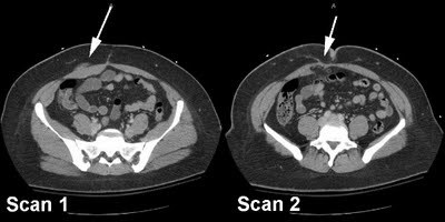
Scan 1 shows blood tracking to the anterior fascia, as well as an increase in size of the rectus muscle.

Scan 2 shows penetration of the posterior rectus sheath with intra-abdominal fat herniating into it. The transverse colon is only 2 cm away deep to it. Scan 1 alone is enough to prompt you to take the patient to the OR!

Can a Normal CT Scan Alone Clear the Cervical Spine in Obtunded Trauma Patients?

This is the first in a series of articles on interesting abstracts presented at the 23rd Annual Scientific Assembly of the Eastern Association for the Surgery of Trauma in Phoenix, Arizona.

C-spine clearance in obtunded trauma patients has been problematic for some time. The options have been:

  • CT plus MRI. This is probably only valid for the first 72 hours after injury, and entails some risk in placing a critically ill patient inside the MRI for 30 minutes or more.
  • CT plus flexion/extension images under fluoroscopy. These are generally only performed by a few brave souls.
  • Leave the collar on until a clinical exam can be performed. This frequently leads to significant skin breakdown problems.

The authors have been reviewing their experience with using CT scan alone. In this paper, they used this technique in patients who met the following criteria:

  • Obtunded
  • Blunt trauma
  • CT normal, as read by a neuroradiologist
  • Moving all extremities

They studied 197 patients, and found no injuries in all surviving patients (11% were lost to followup). One deceased patient had a stable ligamentous injury without spine fracture seen at autopsy. Using this technique resulted in a decrease in the average number of days to spine clearance from 7.5 to 3.3 days, a decrease in skin breakdown from 5% to 0.5%. A decreased length of stay from 23.4 to 13.8 days was also seen, but this could not be attributed to the collar.

Very intriguing! However, the fear of SCIWORA is high in all who clear c-spines. The rarity of this catastrophic problem means that no existing study has the statistical power to show that this type of clearance is safe.

Bottom line: We all need to decide “How many missed injuries is okay?” We will never be able to absolutely clear 100.000% of c-spines by xray alone, or even by adding a clinical exam. This study provides support for one technique, but eventually a catastrophic injury will occur. Who will decide what constitutes an acceptable complication and with what frequency they will occur?

Reference: A Normal CT Alone May Clear the Cervical Spine in Obtunded Blunt Trauma Patients with Gross Extremity Movement – A Prospective Evaluation of a Revised Protocol. Leukhardt, Como, Anderson, Wilczewski, Samia, Claridge. MetroHealth Medical Center, Cleveland, OH.