Tag Archives: chest tube

What The Heck? Pigtail Catheter Chest Tube – The Answer

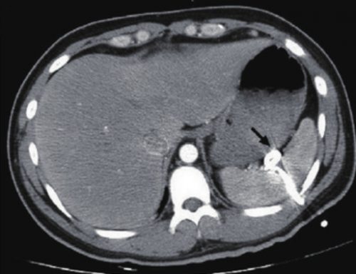
I previously described a trauma patient who had a pigtail type chest tube inserted with some odd CT findings after insertion:

So what is wrong in this picture? Well, the catheter has been inserted into the spleen! This can occur if it is inserted too low, or if there are adhesions between lung and chest wall or diaphragm.

How can it be avoided? Make sure that the insertion point is no lower than the 5th intercostal space. This is the level of the nipple in a male. And depending on what type of kit you use, be careful! Some are based on Seldinger technique, which would seem to be a bit safer. Others use a small trochar, which can be inserted a little too deeply at times. Note that this complication can occur with any kit, and can also occur when using a standard tube and open insertion technique.

Does a pigtail tube even work for hemothorax? There’s some debate about this. Traumatic hemothorax is not defibrinated like a medical one. Thus, there are frequently clots present which may not fully evacuate through a standard chest tube, let alone a tiny one. Thus, I don’t recommend a pigtail for acute traumatic hemothorax.

How should I manage this issue? Obviously, this tube needs to come out. And assuming that the initial indication for the tube is still present, a better one needs to be inserted. Dont’ pull it out yet! First, look at the vital signs. If there is significant bleeding and/or vitals are not normal, an immediate trip to the operating room is in order. In this case, the patient will likely lose their spleen.

If vital signs are stable, book both an interventional radiology suite and an OR. Or better yet, use a hybrid room. Have the radiologist obtain a baseline angiogram, and position a catheter in the main splenic artery. Incrementally remove the pigtail, hand injecting a small amount of contrast each time. If extravasation is noted at any time, the radiologist can then attempt to embolize. If selective embolization isn’t successful, then the main splenic artery should be embolized. If embolization doesn’t work, or vital signs deteriorate at any time, the surgeon should immediately proceed to laparotomy. Attempts at splenic salvage will probably not be successful.

Finally, insert a new, conventional chest tube using finger guidance. Don’t make the same mistake twice! And by the way, this works for pigtails in the liver, too. They are less likely to bleed significantly when withdrawn, and obviously the radiologist can only used selective embolization if they do.

What The Heck? Pigtail Catheter Chest Tube

Here’s a case to make you think!

A patient arrives after being t-boned in his driver side door. He complains of left sided chest and abdominal pain. Chest x-ray shows a modest left hemopneumothorax. The decision is made to insert a pigtail type chest tube, and this is carried out in your trauma bay. It is uneventful, and a small amount of blood but no air is returned. The pelvis x-ray is unremarkable

The patient is then taken to CT, where an abdomen/pelvis scan with contrast is performed. This interesting slice is noted. What the heck?!

Here are my questions:

  • What is wrong in this picture?
  • How could it have been avoided?
  • Does a pigtail chest tube work for hemothorax?
  • How should this issue be managed, and where?

I’ll address these questions in my next post, and more!

Image source: internet

How To: Insert A Small Percutaneous Chest Tube

This short (10 minute) video demonstrated the technique for inserting small chest tubes, also known as “pigtail catheters.” It features Jessie Nelson MD from the Regions Hospital Department of Emergency Medicine. It was first shown at the third annual Trauma Education: The Next Education conference in September 2015, for which she was a course director.

Please feel free to leave any comments or ask any questions that you may have.

YouTube player

Related posts:
Pigtail catheters vs regular chest tubes
Tips for regular chest tubes 

Chest X-Ray After Chest Tube Insertion: Why Do We Do It?

More dogma, or is it actually useful? Any time a chest tube (tube thoracostomy) is inserted, we automatically order a chest x-ray. Even the ATLS course recommends obtaining an image after placement. But anything we do “automatically” is grounds for critical analysis to see if there is a valid reason for doing it.

A South African group looked at the utility of this practice retrospectively in 1004 of their patients. They place 1042 tubes. Here are the factoids:

  • Patients were included if they had at least one chest x-ray obtained after insertion
  • Patients were grouped as follows: Group A (10%) had the tube inserted on clinical grounds with no pre-insertion x-ray (e.g. tension pneumothorax). Group B (19%) had a chest x-ray before and had ongoing clinical concerns after insertion. Group C (71%) had a chest-xray before and no ongoing concerns.
  • 75% of injuries were penetrating (75% stab, 25% GSW), 25% were blunt
  • Group A (insertion with pre-x-ray): 9% had post-insertion findings that prompted a management change (kinked, not inserted far enough)
  • Group B (ongoing clinical concerns): 58% required a management change based on the post-x-ray. 33% were subcutaneous or not inserted far enough (!!)
  • Group C (no ongoing clinical concerns): 32 of 710 (5%) required a management change, usually because the tube was too deep

The authors concluded that if there are no clinical concerns (tube functioning, no clinical symptoms) after insertion, then a chest x-ray is not necessary.

Bottom line: But I disagree with the authors! Even with no obvious clinical concerns, the tube may not be functioning for a variety of reasons. Hopefully, this fact would then be discovered the next day when another x-ray is obtained. But this delays the usual progression toward removing the tube promptly by at least one day. It increases hospital stay, as well as the likelihood of infection or other hospital-associated complication. A chest x-ray is cheap compared to a day in the hospital, which would potentially happen in 5% of these patients. I recommend that we continue to obtain a simple one-view chest x-ray after tube insertion.

Related posts:

Reference: What is the yield of routine chest radiography following tube thoracostomy for trauma?  Injury 46(1):45-48, 2015.