Tag Archives: chest tube

Best of AAST #6: Antibiotics For Chest Tubes??

For as long as I can remember (nearly 50 years worth of literature) there has been some debate about giving antibiotics after chest tube insertion to decrease the infection rate. The pendulum moved back and forth for decades, never getting very far into the “give antibiotics” side. It’s been quite a while since I remember any new papers on this, and I thought the debate had been resolved in favor of never using them.

But then I see an abstract from the AAST multi-institutional trials group studying presumptive antibiotics after chest tube insertion! They conducted a prospective, observational study at 22 Level I trauma centers, enrolling nearly 2,000 patients. They matched patients in antibiotic and no antibiotic groups, arriving at (only) 272 patients in each group.

Here are the results:

Bottom line: First, it’s a little disappointing that the numbers were so low with a trial that includes 22 trauma centers. Did they have a hard time finding centers that would give antibiotics? Or was it just hard to match patients for the variables they were looking at? Regardless, there were no significant differences in infectious complications, and a non-clinically significant difference in ICU stay with antibiotics.

Why won’t this die? If there are so few papers that show an actual benefit from giving antibiotics after chest tube insertion with 50 years of data, then it’s very unlikely that it will ever be shown to be necessary!

Reference: Presumptive antibiotics for tube thoracostomy for traumatic pneumothorax. Session XXII Paper 49, AAST 2018.

Where Did The French Tube Size System Come From?

Medicine sure has some weird measurement systems. Besides the more standardized units like microliters, milligrams, and International Units, we’ve got some odd stuff like French (tubes) and gauge (needles). When dealing with tubes and catheters, the size is usually specified in French units.

Where did the French system come from? It was introduced by a Swiss-born gentleman named Joseph-Frédéric-Benoît Charrière. He moved to Paris and was apprenticed to a knife maker. At the age of 17, he founded a  company that manufactured surgical instruments. His company developed and improved a number of surgical instruments, including hypodermic needles and various catheters.

Charrière introduced the system for describing catheters based on their outer diameter (OD).  It was actually named after him, and in France one will occasionally see catheters described in Ch units. Unfortunately, we Americans had a hard time pronouncing his name, and changed it to the French system (Fr).

So what’s the translation? The Ch or Fr number is the outer diameter of a catheter in millimeters multiplied by 3. It is not the outer circumference in millimeters, and the use of pi is not involved. So a big chest tube (36 Fr) has an OD of 12 mm, and a bigger chest tube (40 Fr) has an OD of 13.33 mm.

Tomorrow: Where did the needle gauge size come from?

Chest Tubes: Size Doesn’t Matter – Part 2

A few days ago, I wrote about a paper that seemed to suggest that using a smaller chest tube (28-32 Fr) vs larger ones (36-40 Fr). The results suggested that their function was very similar. I emphasized that I thought the result was intriguing, because I’m of the opinion that bigger is better for getting clotted blood out. However, I am amenable to changing my mind based on newer, better data.

But I did caution readers that I would like to see more data. One study should never change your practice! Then I see a lot of chatter on Twitter about another study from 2016 that looks at even smaller tubes, with people saying they will now switch to pigtail catheters (12 Fr)!!

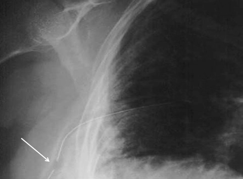
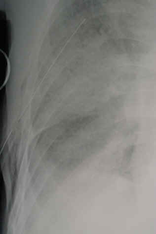
First, not a logical progression of thinking there. And second, let’s take an actual look at the paper. It’s from an emergency medicine group in Fukui, Japan, which retrospectively reviewed their 7 year experience with using a small (20-22 Fr) vs large (28 Fr) tubes. They identified a total of 124 chest tube insertions to compare, 68 small and 56 large.

Now let’s look at the factoids:

  • Demographics, mechanism, and ISS were the same between groups
  • Duration of insertion and initial drainage were also the same between groups
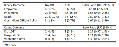
  • Complication rates were similar, with 1 empyema and 2 retained hemothoraces in each group
  • Additional tubes were place in 2 patients with small tubes vs 4 with large tubes
  • Thoracotomy was performed in 2 patients with small tubes vs 1 with a large tube

Based on all of this, the authors concluded that there was no difference in drainage efficacy, complications, or need for additional invasive procedures.

Wait a minute!! Again, if you only read the abstract, you might be led to start using ever smaller chest tubes. But read the entire paper! There are many problems with this paper, including:

  • It’s a very small, retrospective review. This automatically means that the statistical power is suspect.
  • Why did they only document 124 insertions over 7 years?? That’s about one every 3 weeks! Either a lot of data are missing or they are not very busy. But Fukui Prefectural Hospital has over 1000 beds! So it’s the former, not the latter.
  • The retrospective nature means it is not possible to determine why a particular tube size was chosen. Roll of the dice? This fact alone introduces a huge potential for selection bias. Was a smaller tube selected because the hemothorax looked smaller? Probably! The fact that 4 patients with larger tubes had another one placed suggests that they were being used for larger collections. And patients with higher ISS tended to get bigger tubes.

Bottom line: Don’t change your practice based on this paper. And certainly don’t choose to use even smaller pigtails. And of course, always critically read any paper that you like to make sure you are not cherry picking the ones you choose to believe. IMHO, it’s still best to use big (36 Fr) or bigger (40 Fr).

Reference: Small tube thoracostomy (20-22 Fr) in emergent management of chest trauma. Injury 48:1884-1887, 2016.

Chest Tube Size Doesn’t Matter?

It’s great when you read a study that supports your own biases. But it’s not pleasant at all when you find one that refutes what you’ve been teaching for years. Well, I found one of those and I wanted to share it with you.

I’ve always said that there are only two sizes of chest tube for trauma, big (36Fr) and bigger (40Fr). Although there was never any good literature, it seemed intuitive that a large tube would help ensure drainage of bigger clots if hemothorax was present.

A multicenter observational study was carried out that looked at 353 chest tube insertions. This work monitored retained hemothorax or pneumothorax, the need for tube reinsertion or invasive procedure due to incomplete drainage, and pain during insertion.

Here are the factoids:

  • There was roughly a 50:50 large (36-40Fr) vs small (28-32Fr) mix of chest tubes
  • Tubes inserted for hemothorax were also a 50:50 mix of large vs small
  • The initial amount of blood out was small and about the same for both groups
  • There was no significant difference in pneumonia, retained hemothorax, or empyema
  • The need for an invasive procedure (VATS or thoracotomy) was about 11% in both groups
  • Interestingly, there was no difference in visual analog pain score between the groups either.

Bottom line: Basically, large tube and small tube were the same. So maybe chest tube size selection doesn’t matter as much as we (I?) think. It seems to make sense to select a tube size based on your patient’s chest wall, not dogma. Although subjective pain seems to be the same as well, pain and sedation management are key because this is not a fun procedure for the patient, regardless of tube size. I’m not fully convinced yet, and would like to see an additional confirmation study if possible.

Reference: Does size matter? A prospective analysis of 28–32 versus 36–40 French chest tube size in trauma. J Trauma 72(2):422-427, 2012.

What The Heck? Pigtail Catheter Chest Tube – The Answer

I previously described a trauma patient who had a pigtail type chest tube inserted with some odd CT findings after insertion:

So what is wrong in this picture? Well, the catheter has been inserted into the spleen! This can occur if it is inserted too low, or if there are adhesions between lung and chest wall or diaphragm.

How can it be avoided? Make sure that the insertion point is no lower than the 5th intercostal space. This is the level of the nipple in a male. And depending on what type of kit you use, be careful! Some are based on Seldinger technique, which would seem to be a bit safer. Others use a small trochar, which can be inserted a little too deeply at times. Note that this complication can occur with any kit, and can also occur when using a standard tube and open insertion technique.

Does a pigtail tube even work for hemothorax? There’s some debate about this. Traumatic hemothorax is not defibrinated like a medical one. Thus, there are frequently clots present which may not fully evacuate through a standard chest tube, let alone a tiny one. Thus, I don’t recommend a pigtail for acute traumatic hemothorax.

How should I manage this issue? Obviously, this tube needs to come out. And assuming that the initial indication for the tube is still present, a better one needs to be inserted. Dont’ pull it out yet! First, look at the vital signs. If there is significant bleeding and/or vitals are not normal, an immediate trip to the operating room is in order. In this case, the patient will likely lose their spleen.

If vital signs are stable, book both an interventional radiology suite and an OR. Or better yet, use a hybrid room. Have the radiologist obtain a baseline angiogram, and position a catheter in the main splenic artery. Incrementally remove the pigtail, hand injecting a small amount of contrast each time. If extravasation is noted at any time, the radiologist can then attempt to embolize. If selective embolization isn’t successful, then the main splenic artery should be embolized. If embolization doesn’t work, or vital signs deteriorate at any time, the surgeon should immediately proceed to laparotomy. Attempts at splenic salvage will probably not be successful.

Finally, insert a new, conventional chest tube using finger guidance. Don’t make the same mistake twice! And by the way, this works for pigtails in the liver, too. They are less likely to bleed significantly when withdrawn, and obviously the radiologist can only used selective embolization if they do.