Tag Archives: Cervical spine

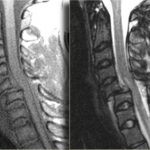
Clearing The Cervical Spine With MRI

If you follow the trauma literature, clearance of the cervical spine in obtunded patients is confusing at best. Although there is some literature out there that suggests that a good cervical CT alone is adequate, I’m not a believer. I’ve seen a case where the radiologist called the scan normal and a good spine surgeon called an injury and was right. So I’m reluctant to use CT alone because the skills of radiologists vary widely. I might be able to believe a dedicated neuroradiologist, but you can’t guarantee one will be reading your patient’s images.

So I fall back on the routine of clearing the bones with a CT scan, and the ligaments with something else. That something else could be a clinical exam (not available in the obtunded patient), flexion-extension images under fluoroscopy (makes a lot of people nervous), keeping the patient in a collar for weeks (skin breakdown), or an MRI. The problem is that there is little guidance in the literature regarding how good MRI is or the best way to use it.

A recent paper in the Journal of Trauma retrospectively looked at 512 out of 17,000 patients (!) seen over 5 years at one trauma center who had both CT and MRI of the c-spine. They wanted to determine if MRI was of any value in cervical spine clearance. Only 150 met the inclusion criteria (GCS<13, no obvious neuro deficit, normal CT). Half of the MRIs were normal. Of the abnormal ones, 81% showed a ligamentous or soft tissue injury. None were deemed unstable and no specific management was needed for any of the abnormal scans.

The authors interpreted their data as showing that MRI provided no additional useful information. However, numbers were (very) small, so the likelihood of them seeing someone with an unstable ligamentous injury was low. Could it be that they showed that MRI detected stable injuries well, and that they could essentially remove the collar based on that?

Bottom line: We still don’t know how to use MRI for clearance. My bias (no good data I can find) is that it is good in suggesting ligamentous injury via nearby edema. If this injury involves only one set of ligaments, it is very likely a stable one and the collar can be removed. If it involves several groups of ligaments, that is probably not the case. And how soon do we have to get the MRI after injury? Some have suggested that 72 hours is the ideal window because edema decreases afterwards. Sounds reasonable, but I can’t find a shred of evidence in the literature. For now, I’ll get an MRI within 72 hours and if it is abnormal, pass the buck to my neurosurgical colleagues so they can gnash their teeth, too.

I would be very happy if someone can help me out and point me towards some good literature on this topic!

Reference: The value of cervical magnetic resonance imaging in the evaluation of the obtunded or comatose patient with cervical trauma, no other abnormal neurological findings, and a normal cervical computed tomography. J Trauma 72(3):699-702, 2012.

More on Distracting Injury and Spine Clearance

There is a lot of angst out there among trauma professionals when it comes to clearing the cervical spine and possible distracting injuries. I’ve written about this before, and the most important technique I use is to try to see if the patient is aware of pain in areas distant from the suspected distraction.

A recent prospective study looked at injury patterns and c-spine clearance in over 9,000 trauma patients at a single Level I trauma center. Of those, 101 were evaluable (not intoxicated, no head injury) and actually had a cervical fracture. 96% of them were symptomatic, despite the majority having what would normally be considered a distracting injury (87%). Of the 4 who did not have pain or tenderness on examination of the neck, every one of them had a distraction.

There was a pattern as to which “distracting” injuries were really a distraction. All four of the asymptomatic patients had bruising or tenderness to the upper anterior chest, which diverted their attention away from their neck. Other injuries caused pain in some of these patients, but it was outweighed by the chest wall pain.

Bottom line: Distracting injury is currently defined too liberally, which results in lots of patients getting lots of unneeded cervical spine imaging. Although this study is small, it adds one more piece of information to the c-spine clearance puzzle. I personally will add this to my current practice and clear the cervical spine if:

  • The patient is aware of multiple sources of pain
  • Subjective pain scale overall is less than 6 (otherwise provide better pain control!)
  • There is no pain/tenderness/injury to the upper chest 

Related posts:

Reference: The presence of nonthoracic distracting injuries does not affect the initial clinical examination of the cervical spine in evaluable blunt trauma patients: a prospective observational study. J Trauma 71(3):528-532, 2011.

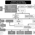
Algorithm For Clearing the Pediatric Cervical Spine

I previously wrote about a straightforward way to clear the cervical spine in children. Click here to see the article. Alfred I. DuPont Children’s Hospital has condensed their clearance technique into a relatively simple algorithm that can be used in conjunction with my previous tips.

Some notes on this algorithm:

  • Can be performed only by attending physicians or a trauma resident in consultation with the attending trauma surgeon
  • Clinical clearance alone may be carried out in select cases
  • If radiographs are required, cross-table lateral, anterior/posterior, and odontoid views should be obtained (age 8 and above, non-intubated)
  • Flexion / extension views should only be ordered in consultation with neurosurgery

Download a print version of the protocol here

Related post: How Do I Clear The Pediatric Cervical Spine?

Image and protocol courtesy of the Alfred I DuPont Children’s Hospital

Comparison of Cervical Spine Stabilization

Eight months ago I blogged about inline stabilization vs inline traction of the cervical spine. Click here to read the post. A reader recently asked what the optimal method for inline stabilization is.

We’ve been pondering this question for nearly 30 years. In 1983, trauma surgeons at UCLA looked at a number of devices available at that time and tested them on normal volunteers. They measured neck motion to see which was “best." 

Here’s what they found:

  • Soft collar – In general, this decreased rotation by 8 degrees but insignificantly protected against flexion and extension. Basically, this keeps your neck warm and little else.
  • Hard collars – A variety of collars available in that era were tested. They all allowed about 8% flexion, 18% lateral movement, and 2% rotation. The Philadelphia collar allowed the least extension.
  • Sandbags and tape – Surprisingly, this was the best. It allowed no flexion and only a few percent movement in any other direction.

The Mayo clinic compared four specific hard collars in 2007 (Miami J, Miami J with Occian back, Aspen, Philadelphia). They found that the Miami J and Philadelphia collars reduced neck movement the best. The Miami J with or without the Occian back provided the best relief from pressure. The Aspen allowed more movement in all axes.

And finally, the halo vest is the gold standard. These tend to be used rarely and in very special circumstances.

Bottom line: 

  • For EMS: Rigid collar per your protocol is the standard. In a pinch you can use good old tape and sandbags with excellent results.
  • For physicians: The Miami J provides the most limitation of movement. If the collar will be needed for more than a short time, consider the well-padded Occian back Miami J (see below).

Miami J with Occian back

References:

  • Efficacy of cervical spine immobilization methods. J Trauma 23(6):461-465, 1983.
  • Range-of-motion restriction and craniofacial tissue-interface pressure from four cervical collars. J Trauma 63(5):1120, 1126, 2007.

From the Archives: Cervical Spine Imaging

So far, I’ve posted more than 200 items during the past year and a half. There’s a lot of good stuff in the archives, and I am going to periodically provide a list of links to them so they are not forgotten. To see a full index of the archive by subject, click here.

Today, I’m going to focus on cervical spine imaging. Here are four interesting posts from the archives:

As always, I welcome suggestions for new posts!