Tag Archives: Cervical spine

Cervical Spinal Cord Injury: Who Needs A Tracheostomy?

The sad truth is that patients with cervical spine injury may need a tracheostomy. In very high lesions (C1-2) the need may be permanent. Lower injuries (C3-5) frequently need a trach for a limited period of time while they develop enough reserve to compensate for the lost of chest wall muscle power.

It’s not always easy to tell which patient is likely to need intubation upon arrival in the ED. I’ve seen occasional patients fail while getting their CT scans, which is poor planning. Is there a way to predict who might fail, thus benefiting from early intubation and an early plan for tracheostomy?

The trauma group at LAC + USC Medical Center undertook a National Trauma Databank review to try to answer this question. They identified 5256 patients with cervical spinal cord injuries without a severe traumatic brain injury that would otherwise require intubation. About 21% received tracheostomies, and the common predictors were:

  • Intubation at the scene by EMS (they’ve done the job of deciding for us!)
  • Intubation in the ED
  • Complete cord injury at any level
  • Facial fractures
  • Chest trauma
  • Injury Severity Score >=16

Patients who received a tracheostomy generally spent more days on the vent, in the ICU and in the hospital than those who did not. However, their mortality was lower.

It’s generally recognized that patients with complete injuries from C1-C5 routinely require tracheostomy. The surprising thing about this study was that complete injuries at C6 or C7 did as well.

Bottom line: If you have a patient with a spinal cord injury who meets any of the criteria above, stand ready to intubate. I tell my trainees that, if at any time they see something that makes them think about intubating, they should have already done it. Likewise, the surgical ICU team should have a low threshold to performing an early tracheostomy on these patients.

Reference: Incidence of clinical predictors for tracheostomy after cervical spinal cord injury: a National Trauma Databank review. J Trauma 70(1): 111-115, 2011.

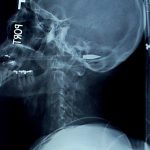
Picture: crossbow bolt through the mouth and cervical spinal cord.

Dysphagia and Cervical Spine Injury

Cervical spine injury presents a host of problems, but one of the least appreciated ones is dysphagia. Many clinicians don’t even think of it, but it is a relatively common problem, especially in the elderly. Swallowing difficulties may arise for several reasons:

  • Prevertebral soft tissue swelling may occur with high cervical spine injuries, leading to changes in the architecture of the posterior pharynx
  • Rigid cervical collars, such as the Miami J and Aspen, and halo vests all force the neck into a neutral position. Elderly patients may have a natural kyphosis, and this change in positioning may interfere with swallowing. Try extending your neck by about 30 degrees and see how much more difficult it is to swallow.
  • Patients with cervical fractures more commonly need a tracheostomy for ventilatory support and/or have a head injury, and these are well known culprits in dysphagia

A study in the Jan 2011 Journal of Trauma outlines the dysphagia problem seen with placement of a halo vest. They studied a series of 79 of their patients who were treated with a halo. A full 66% had problems with their swallowing evaluation. This problem was associated with a significantly longer ICU stay and a somewhat longer overall hospital stay.

Bottom line: Suspect dysphagia in all patients with cervical fractures, especially the elderly. Carry out a formal swallowing evaluation, and adjust the collar or halo if appropriate. 

Reference: Swallowing dysfunction in trauma patients with cervical spine fractures treated with halo-vest fixation. J Trauma 70(1):46-50, 2011.

Distracting Injury and Cervical Spine Clearance

One of the tenets of clinical c-spine clearance is that there be no “distracting injury.” What does this mean exactly? Can the clinician adequately judge which injuries are too distracting?

The Loyola group prospectively looked at 160 patients needing c-spine clearance over a 9 month period. GCS had to be 14 or 15, and the patients were excluded if they were intoxicated or received an analgesic prior to the clearance attempt. A total of 84% had no neck pain, and 82% of those had no peripheral, potentially distracting pain. Patients with perceived distracting pain and those without had very similar Visual Analog Scores (VAS) for pain. 

Overall, the majority of patients and physicians did not believe that distracting pain was present, and when pain was present there was little agreement whether it was distracting. The few patients who did have spine fractures had a VAS for pain >5. The use of physician judgment for distracting pain and clearance worked just fine in this study.

Bottom line: The authors recommend using clinician judgment as to the degree of distracting pain when clearing the c-spine. If you want to be more objective, if the patient complains of a Visual Analog Score for pain of more than 5, then you may want to delay clearance. Note: this is a small study that really needs to be replicated before widespread use.

Reference: C-spine clearance: don’t be distracted – just trust your judgment. Presented at the 24th annual scientific assembly of EAST, Session II, Paper 9. Click here to see the abstract. 

Cervical Spine Clearance and Altered Mental Status

Clearance of the cervical spine is a complicated topic, with many opinions and anecdotes. EAST developed a set of practice guidelines in 1998 and updated them in 2000 and again in 2008. They are well-accepted and very helpful.

Spine clearance in an obtunded or intoxicated patient is made even more challenging. Here’s an approach based on the EAST guidelines that I find helpful:

  1. Clear the bones. Obtain a CT of the cervical spine from skull base to T2. Sagittal and coronal 2D reconstructions must be created for review. Conventional images (AP, lateral, odontoid) are of no additional value.
  2. If a fracture is identified, consult your spine service.
  3. If a neurologic deficit is present, obtain an MRI and consult your neurosurgery or spine service.
  4. Clear the ligaments. In the obtunded patient, there are 3 choices: 1) keep the collar on until the patient wakes up enough to be examined, 2) obtain an MRI to evaluate the ligaments, or 3) remove the collar on the basis of CT alone.

In patients that you don’t expect to wake up any time soon, I prefer MRI. Some say that it should be obtained within 72 hours of injury for best accuracy in detecting ligamentous injury. Unfortunately, I have not been able to find any specific literature support for this. If the MRI is negative, the collar can be removed immediately.

There is a growing body of research that suggests that CT alone is sufficient for clearance. My opinion is that this is probably true, but only if the scan is read by a radiologist who is especially skilled in reading CT scans of the cervical spine. A pool radiologist may miss subtle findings that might indicate a ligamentous injury.

Reference: Eastern Association for the Surgery of Trauma practice guideline: Identifying Cervical Spine Injuries Following Trauma – 2009 Update. Click here to download.

How To: Flexion / Extension Views of the Cervical Spine

Clearance of the cervical spine can often be done using clinical criteria alone (see this video at http://youtu.be/NhjF9kDOcjE). If this is not possible, a combination of radiologic and clinical evaluation is usually carried out. 

In some cases, radiographic studies (usually CT) are normal, but there is pain on clinical exam. Our next step is to send the patient to xray for flexion and extension views. This exam is performed by removing the collar while the patient is sitting, so the thoracic and lumbar spines must be clear before ordering this. The patient then gently flexes and extends the neck to their limits of comfort. Images are then obtained at the limits of flexion and extension. The premise is that a normal, awake patient cannot and will not move their neck beyond their comfort level to the point where they could cause themselves neurologic injury. 

It is very important that you look at the images yourself. The radiologist may review the images and will report that “there is no evidence of subluxation at the limits of flexion and extension.” But the patient may have barely moved their neck!

The question is: how much flexion and extension do you need to have to clear the spine?

The answer is not easy to find, and is buried in literature from the 1980s and 90s. According to the EAST guidelines, the ideal amount is 30 degrees from neutral for both flexion and extension. This is not always achievable in elderly patients, so in those cases you must use your judgment. Talk to the patient to find out if they stopped moving their neck forward or backward due to pain, or because they just can’t move it that far.

Trouble signs to look for are:

  • Subluxation of more that 2mm at any level
  • Angulation of more than 11 degrees

Any abnormality should prompt a spine consult.

If the study is not abnormal but the amount of flexion and/or extension is not adequate, there are two options. First, just leave the collar in place and try again in a week or so and try again. This will allow any soft tissue injuries to get better and may allow a successful repeat study. The alternative is a more costly and less well-tolerated MRI.

References:

  1. EAST Practice Guidelines, Identifying Cervical Spine Injuries Following Trauma – Update (2000).
  2. Defining radiographic criteria for flexion-extension studies of the cervical spine. Robert Knopp et al. Ann Emerg Med. 2001 Jul;38(1):31-5.